Transcription of PROTEINS: THREE-DIMENSIONAL STRUCTURE
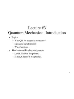
1 CHAPTER 6 PROTEINS: three -DIMENSIONALSTRUCTURE1. secondary STRUCTUREA. The Peptide GroupB. Regular secondary STRUCTURE : The aHelix and thebSheetC. Fibrous ProteinsD. Nonrepetitive protein Structure2. tertiary STRUCTUREA. Determining protein StructureB. Motifs (Supersecondary Structures) and DomainsC. protein Families3. quaternary STRUCTURE AND SYMMETRY4. protein FOLDING AND STABILITYA. Forces That Stabilize protein StructureB. protein Denaturation and RenaturationC. protein Folding PathwaysD. protein Dynamics124 The atomic STRUCTURE of myoglobin, an oxygen binding protein , is drawn here as a stick model. The overall conformation of aprotein such as myoglobin is a function of its amino acid sequence. How do noncovalent forces act on a polypeptide chain tostabilize its unique THREE-DIMENSIONAL arrangement of atoms? [Figure copyrighted by Irving Geis.]For many years, it was thought that proteins were colloids of random struc-ture and that the enzymatic activities of certain crystallized proteins weredue to unknown entities associated with an inert protein carrier.
2 In 1934, Bernal and Dorothy Crowfoot Hodgkin showed that a crystal of theprotein pepsinyielded a discrete diffraction pattern when placed in an X-ray beam. This result provided the first evidence that pepsin was not arandom colloid but an ordered array of atoms organized into a large yetuniquely structured relatively small proteins contain thousands of atoms, almost all of which occupy definite positions in space. The first X-ray STRUCTURE of aprotein, that of sperm whale myoglobin, was reported in 1958 by JohnKendrew and co-workers. At the time only 5 years after James Watsonand Francis Crick had elucidated the simple and elegant STRUCTURE of DNA(Section 3-2B) protein chemists were chagrined by the complexity andapparent lack of regularity in the STRUCTURE of myoglobin.
3 In retrospect,such irregularity seems essential for proteins to fulfill their diverse biolog-ical roles. However, comparisons of the ,7000 protein structures nowknown have revealed that proteins actually exhibit a remarkable degree ofstructural we saw in Section 5-1, the primary STRUCTURE of a protein is its linearsequence of amino acids. In discussing protein STRUCTURE , three further lev-els of structural complexity are customarily invoked: secondary structureis the local spatial arrangement of a polypeptide sbackbone atoms without regard to the conformations of its side chains. tertiary structurerefers to the THREE-DIMENSIONAL STRUCTURE of an en-tire polypeptide. Many proteins are composed of two or more polypeptide chains,loosely referred to as subunits. A protein s quaternary structurerefersto the spatial arrangement of its four levels of protein STRUCTURE are summarized in Fig.
4 6-1. secondary Structure125 Figure of protein STRUCTURE .(a) primary STRUCTURE , (b) secondary STRUCTURE , (c) tertiary STRUCTURE , and (d) quaternary STRUCTURE .[Figure copyrighted by Irving Geis.]Secondarystructure(helix) primary STRUCTURE (amino acid sequence in a polypeptide chain) quaternary STRUCTURE :the four separate chainsof hemoglobin assembledinto an oligomeric proteinTertiary STRUCTURE :one complete protein chain( chain of hemoglobin) 2211(b)(a)(c)(d) Lys Ala His Gly Lys Lys Val Leu Gly - Ala In this chapter, we explore secondary through quaternary STRUCTURE , in-cluding examples of proteins that illustrate each of these levels. We also in-troduce methods for determining THREE-DIMENSIONAL molecular structureand discuss the forces that stabilize folded secondary STRUCTUREP rotein secondary STRUCTURE includes the regular polypeptide folding pat-terns such as helices, sheets, and turns.
5 However, before we discuss thesebasic structural elements, we must consider the geometric properties ofpeptide groups, which underlie all higher order The Peptide GroupIn the 1930s and 1940s, Linus Pauling and Robert Corey determined theX-ray structures of several amino acids and dipeptides in an effort to elu-cidate the conformational constraints on a polypeptide chain. These stud-ies indicated that the peptide group has a rigid, planar STRUCTURE as a conse-quence of resonance interactions that give the peptide bond ,40%double-bond character:This explanation is supported by the observations that a peptide group sCON bond is shorter than its NOCasingle bond and that its CPObond is longer than that of aldehydes and ketones. The planar con-formation maximizes p-bonding overlap, which accounts for the peptidegroup s groups, with few exceptions, assume the trans conformation, inwhich successive Caatoms are on opposite sides of the peptide bond join-ing them (Fig.)
6 6-2). The cis conformation,in which successive Caatoms areon the same side of the peptide bond, is ,8 kJ?mol21less stable than thetrans conformation because of steric interference between neighboring sidechains. However, this steric interference is reduced in peptide bonds to Proresidues, so ,10% of the Pro residues in proteins follow a cis peptide Angles between Peptide Groups Describe Polypeptide Chain ConformationsThe backboneor main chainof a protein refers to the atoms that par-ticipate in peptide bonds, ignoring the side chains of the amino acidCONHCO2NH+126 Chapter 6. Proteins: THREE-DIMENSIONAL 116 C C Peptide 122 NHHOCRRHF igure trans peptide bondlengths (in angstroms) and angles (in degrees) arederived from X-ray crystal structures. [AfterMarsh, and Donohue, J., Adv. protein ,249 (1967).] See Kinemage Exercise chainSide chainFigure conformation of a backbone is shown asa series of planar peptide groups.
7 [Figure copyrighted by Irving Geis.]residues. The backbone can be drawn as a linked sequence of rigid planarpeptide groups (Fig. 6-3). The conformation of the backbone can thereforebe described by the torsion angles(also called dihedral anglesor rotationangles) around the CaON bond (f) and the CaOC bond (c) of each residue(Fig. 6-4). These angles, fand c, are both defined as 180 when the polypep-tide chain is in its fully extended conformation and increase clockwise whenviewed from conformational freedom and therefore the torsion angles of apolypeptide backbone are sterically constrained. Rotation around theCaON and CaOC bonds to form certain combinations of fand canglesmay cause the amide hydrogen, the carbonyl oxygen, or the substituents ofCaof adjacent residues to collide ( , Fig. 6-5). Certain conformations oflonger polypeptides can similarly produce collisions between residues thatare far apart in 6-1.
8 secondary Structure127 Figure angles of the polypeptide planar peptidegroups are shown. The only reasonably free movements are rotations around theCaON bond (measured as f) and the CaOC bond (measured as c). By convention,both fand care 180 in the conformation shown and increase, as indicated, in theclockwise direction when viewed from Ca. [Figure copyrighted by Irving Geis.] Figure interference between adja-cent peptide can result in aconformation in which the amide hydrogen ofone residue and the carbonyl oxygen of the nextare closer than their van der Waals distance. [Figure copyrighted by Irving Geis.] See Kinemage Exercise Ramachandran Diagram Indicates Allowed Conformations of PolypeptidesThe sterically allowed values of fand ccan be calculated. Sterically for-bidden conformations, such as the one shown in Fig. 6-5, have fand cval-ues that would bring atoms closer than the corresponding van der Waalsdistance (the distance of closest contact between nonbonded atoms).
9 Suchinformation is summarized in a Ramachandran diagram(Fig. 6-6), whichis named after its inventor, G. N. areas of the Ramachandran diagram (most combinations of fandc) represent forbidden conformations of a polypeptide chain. Only threesmall regions of the diagram are physically accessible to most residues. Theobserved fand cvalues of accurately determined structures nearly alwaysfall within these allowed regions of the Ramachandran plot. There are,however, some notable cyclic side chain of Pro limits its range of fvalues to angles ofaround 260 , making it, not surprisingly, the most conformationallyrestricted amino acid , the only residue without a Cbatom, is much less sterically hin-dered than the other amino acid residues. Hence, its permissible rangeof fand ccovers a larger area of the Ramachandran diagram.
10 AtGly residues, polypeptide chains often assume conformations that areforbidden to other Regular secondary STRUCTURE : The aHelix and the bSheetA few elements of protein secondary STRUCTURE are so widespread that theyare immediately recognizable in proteins with widely differing amino acidsequences. Both the ahelixand the bsheetare such elements; they are128 Chapter 6. Proteins: THREE-DIMENSIONAL Structure180900 90C 180 180 90090180 (deg) (deg) LFigure Ramachandran green-shaded regions indicate thesterically allowed fand cangles for all residues except Gly and Pro. The orangecircles represent conformational angles of several secondary structures: a, right-handed ahelix; hh, parallel bsheet; hg, antiparallel bsheet; C, collagen helix; aL,left-handed regular secondary structures because they are composed of se-quences of residues with repeating fand aHelixOnly one polypeptide helix has both a favorable hydrogen bonding pat-tern and fand cvalues that fall within the fully allowed regions of theRamachandran diagram: the ahelix.