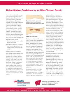
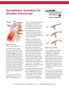
Transcription of Rehabilitation Guidelines for Meniscal Repair of Root and ...
1 UW HEALTH SPORTS REHABILITATIONThe world class health care team for the UW Badgers and proud sponsor of UW 621 SCIENCE DRIVE MADISON, WI 53711 4602 EASTPARK BLVD. MADISON, WI 53718 Rehabilitation Guidelines for Meniscal Repair of root and Complex TearsThere are two types of cartilage in the knee; articular cartilage and meniscus cartilage. Articular cartilage is made up of collagen, proteoglycans and water which lines the end of the bones that meet to form a joint. The primary function of the articular cartilage is to provide a smooth gliding surface for joint motion.
2 Rubbing articular cartilage on articular cartilage is approximately 5 times more smooth, or with less friction, than rubbing ice on meniscus cartilage in the knee includes a medial (inside) meniscus and a lateral (outside) meniscus. Together they are referred to as menisci. The menisci are wedge shaped, being thinner toward the center of the knee and thicker toward the outside of the knee joint (Figures 1 3). This shape is very important to its function. The primary function of the menisci is to improve load transmission. A relatively round femur sitting on a relatively flat tibia forms the knee joint.
3 Without the menisci the area of contact force between these two bones would be relatively small, increasing the contact stress by 235-335% (Figure 4). The menisci also provide some shock absorption, lubrication and joint stability. The menisci are composed of an anterior horn, body and posterior horn, with each horn anchored to the tibia by a strong anterior and posterior root . The posterior roots provide secondary stability to the knee LateralmeniscusMedialmeniscusFigure 1 Meniscus cartilage (shown here from above the knee, without the femur) Image property of Primal Pictures, Ltd.
4 , Use of this image without authorization from Primal Pictures, Ltd. is 2 Medial (inside) view of the kneeImage property of Primal Pictures, Ltd., Use of this image without authorization from Primal Pictures, Ltd. is 3 Normal MRI (saggital view) of the knee, lateral (outside) viewFigure 4 Schematic representation of the Meniscal effect on contact pressure in the knee. Contact area is increased by 50% with addition of menisci. This reduces contact meniscuswith meniscusRehabilitation Guidelines For Meniscal Repair for root 621 SCIENCE DRIVE MADISON, WI 53711 4602 EASTPARK BLVD.
5 MADISON, WI 53718and support against tibial rotation. The function of these are extremely important in protecting the articular cartilage (Figure 5). Studies have shown that Meniscal root tears have a high incidence of recurrent Meniscal injury and stress, which continues to weaken the Meniscal tissue over process of tissue degeneration makes it very unlikely that a surgical Repair will heal or that the surrounding meniscus will be strong enough to hold the sutures used to Repair it. One report showed that less than 10% of Meniscal tears occurring in patients greater than 40 years of age were repairable.
6 Symptoms of a degenerative meniscus tear include swelling, pain along the joint line, catching and locking. Most often degenerative tears are surgically removed. Occasionally a patient may be able to regain function through Rehabilitation without surgery progressing to knee arthritis. There are two categories of Meniscal tears: acute traumatic tears and degenerative tears. Degenerative tears most commonly occur in middle-aged people. Acute traumatic tears occur most frequently in the athletic population as a result of a twisting injury to the knee when the foot is planted. Symptoms of an acute meniscus tear include swelling, pain along the joint line, catching, locking and a specific injury.
7 Often times these tears can be diagnosed by taking a thorough history and completing a physical examination. An MRI may be used to assist in making the an athlete suffers a Meniscal root tear, the three options for treatment include: non-operative Rehabilitation , surgery to trim out the area of torn meniscus or surgery to Repair (stitch together) the torn meniscus. root tears are repaired whenever possible based on their importance in limiting future knee arthritis. Repair can be achieved by a transtibial tunnel Repair , a suture anchor Repair or a side to side Repair . Figure 6 shows the blood vessels (perimeniscular capillary plexus) entering the outer portion of the meniscus.
8 This blood supply is necessary for a tear or a Repair to heal. After Meniscal surgery, Rehabilitation with a physical therapist or athletic trainer is needed to restore range of motion, strength, movement control and guide the athlete s return to sport. When the meniscus root is repaired there will be a prolonged period of restricted weight bearing and range of motion to protect the healing tissue. The following Rehabilitation Guidelines are presented in a criterion based progression. Specific time frames, restrictions and precautions are given to protect healing tissues and the surgical Repair /reconstruction.
9 General time frames are also given for reference to the average individual, but individual patients will progress at different rates depending on their age, associated injuries, pre-injury health status, Rehabilitation compliance and injury severity. The size and location of the Meniscal tear may also affect the rate of post-operative 5 MRI images of an intact and torn Meniscal root . Image #1 shows an intact medial meniscus root connecting to the tibia. Image #2 shows a torn medial Meniscal root . Figure 6 Perimeniscular capillary plexus (thick arrow) providing blood supply to the outer third of the Guidelines For Meniscal Repair for root 621 SCIENCE DRIVE MADISON, WI 53711 4602 EASTPARK BLVD.
10 MADISON, WI 621 SCIENCE DRIVE MADISON, WI 53711 4602 EASTPARK BLVD. MADISON, WI 53718 PHASE I (Surgery to 8 weeks after surgery)Appointments Rehabilitation appointments begin 3-5 days post-operatively and then approximately 1 time per week Rehabilitation Goals Protection of the post-surgical knee Eliminate effusion (swelling) Restore leg controlPrecautions Weightbearing: touch down weight bearing (TDWB) with crutches Brace: post-operative extension brace for 6 weeks. Wean from brace locked to unlocked to no brace after 6 weeks and as patient establishes leg control, pain control and safe gait mechanics.