Transcription of Rehabilitation Guidelines For Periacetabular Osteotomy ...
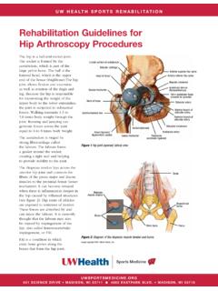
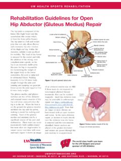
1 Rehabilitation Guidelines For Periacetabular Osteotomy (PAO) Of The HipThe hip joint is composed of the femur (the thigh bone) and the acetabulum (the socket formed by the three pelvic bones). The hip joint is a ball and socket joint that not only allows flexion and extension, but also rotation of the thigh and leg (Fig 1). The head of the femur is encased by the bony socket in addition to a strong, non-compliant joint capsule, making the hip an extremely stable joint. Because the hip is responsible for transmitting the weight of the upper body to the lower extremities and the forces of weight bearing from the foot back up through the pelvis, the joint is subjected to substantial forces (Fig 2). Walking transmits to times body weight through the joint and running and jumping can generate forces across the joint equal to 6 to 8 times body weight.
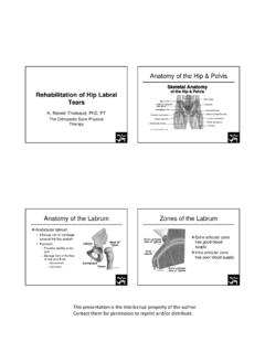
2 The labrum is a circular, fibrocartilaginous structure that surrounds the socket. It functions to seal the joint, enhance stability and provide proprioceptive feedback (a sense of joint position) to the brain and central nervous system. The labrum acts as a suction seal or gasket for the hip joint. This helps to maintain the hydrostatic pressure that protects the articular cartilage on the head of the femur and the the acetabulum (socket) does not fully form, the result can be hip dysplasia. This causes the hip joint to experience load that is poorly tolerated over time, resulting in joint pain and restricted movement. Note the difference of how far the socket covers the head of the femur in Figure 3 compared to Figure 4 below. While this type of joint abnormality is usually present from birth, patients often do not become symptomatic until adolescence or early adulthood.
3 An adequate amount of acetabular (socket) depth creates load distribution that is shared by the whole hip, including joint surfaces and the previously-mentioned acetabular dysplasia is present, loads transferred through the hip joint can be more focal and result in overload of cartilage and bone causing injury that can progress to osteoarthritis. This condition can occur with femoroacetabular impingement and labral the symptomatic dysplasia of the hip is not caught soon enough and progresses to arthritis, the patient would be more of a candidate for Lunate surface of acetabulumArticular cartilageHead of femurNeck of femurIschial tuberosityGreater trochanterLesser trochanterTransverseacetabular ligamentAcetabular arteryObturator membranePosterior branch of obturator arteryObturator arteryFat in acetabular fossa(covered by synovial)Acetabular labrum(fibrocartilainous)Iliopubic eminenceAnterior inferior iliac spineAnterior superior iliac spineAnterior branch of obturator arteryIntertrochanteric lineRound ligament(ligamentum capitis)Figure 1 Hip joint (opened)
4 Lateral view UW HEALTH SPORTS REHABILITATIONThe world class health care team for the UW Badgers and proud sponsor of UW 621 SCIENCE DRIVE MADISON, WI 53711 4602 EASTPARK BLVD. MADISON, WI 53718 Rehabilitation Guidelines For Periacetabular Osteotomy (PAO) Of The Hip 2a total hip replacement. If arthritic changes are minimal, or absent, the patient might be a candidate for a Periacetabular Osteotomy (PAO). A PAO is an open procedure where the socket is separated from the rest of the pelvis by making three cuts in the pelvis. It is then repositioned to better cover the femoral head and secured with long screws and potentially bone grafting material (Fig 5 and 6). This surgery will require a short stay in the a torn labrum is present, this can be addressed with a hip arthroscopy prior to the PAO portion of the surgery (see for more details).
5 If cam type femoroacetabular impingement is present (FAI), it can be addressed after the PAO is of the information gathering in determining if a patient might benefit from a PAO, with or without hip arthroscopy, includes imaging. Radiographs, or x-rays, give a good initial view of bony alignment and help diagnose the hip dysplasia. Magnetic resonance imaging, or MRI, shows the soft tissue such as the labrum or cartilage that covers the bony surfaces of the hip joint. A hip CT scan provides excellent three- dimensional anatomy of the pelvis and femur for surgical planning and a better understanding of hip joint surgeon may recommend anesthetic or corticosteroid injections to treat pain and help identify the origin of your pain which helps to determine if surgery could be helpful. Physical therapy with a provider who is experienced in treating hip conditions both pre-and post-operatively is usually recommended as this can help patients avoid surgery or strengthen their hip and core muscles to make the post-surgical recovery a little bit surgery, a patient spends 2-5 days in the hospital.
6 They will learn to walk with crutches or a walker, usually about day 2 after surgery, minimizing weight bearing on the leg until the newly positioned socket heals. They will have to maintain this partial weight bearing status for about 6 the patient will begin outpatient physical therapy at about 3 weeks eventually being able to return to full weight bearing, walking without crutches and even athletics. Physical therapy will help strengthen the muscles around the hip and pelvis, restore range of motion of the hip joint and integrate the newly aligned hip into a patient s overall daily 2: Image depicting force transmittal through the hip 621 SCIENCE DRIVE MADISON, WI 53711 4602 EASTPARK BLVD. MADISON, WI 53718 Figure 3: Pelvic radiograph with measurements of the lateral center edge angle (LCEA) on the patient s left hip.
7 Normal LCEA is >25 . This patient would be diagnosed with hip dysplasia. Figure 4: Pelvic radiograph with measurements of the lateral center edge angle (LCEA) on the patient s left hip. Normal LCEA is >25 . 17 25 Rehabilitation Guidelines For Periacetabular Osteotomy (PAO) Of The Hip 3 The Rehabilitation Guidelines are presented in a criterion-based progression. The patient may also have postoperative hip and thigh pain and numbness of the groin, thigh, and/or pelvis near and around incision but these symptoms usually resolve over Rehabilitation Principals:1. Post-operative recovery begins with preoperative Rehabilitation , preoperative hip and core strengthening, review of gait training with assistive device and discussion of post-operative equipment needs; elevated toilet seat, wheelchair and long handled reachers are A continuous passive motion (CPM) machine will be issued at the preoperative workup appointment and will be used with a setting of 0 degrees extension and 30 degrees of flexion for eight hours per day until the first post-operative appointment at 2-3 weeks.
8 After the first week, the flexion range of motion can be gradually increased up to 90 degrees if the patient remains Patients will be limited in weightbearing for the first 6 weeks. No more than 20 pounds of body weight and crutches or a walker will need to be used for all walking. Patients should avoid prolonged sitting or lying on the surgical side for the first 4-6 5 (multiple figures above): These images demonstrate the cuts made around the acetabulum and how they are repositioned. Image copyright 2018: UW Health Sports Medicine. 621 SCIENCE DRIVE MADISON, WI 53711 4602 EASTPARK BLVD. MADISON, WI 621 SCIENCE DRIVE MADISON, WI 53711 4602 EASTPARK BLVD. MADISON, WI 53718 Figure 6: The before (left) and after (right) pelvis radiographs of a patient who underwent PAO. Notice the increased coverage indicated by the yellow arrow after Guidelines For Periacetabular Osteotomy (PAO) Of The Hip 4 PHASE I (Surgery to 6 weeks)
9 Appointments Surgery will require an inpatient hospital stay of 2-5 days Inpatient Rehabilitation begins post-op day 1, with emphasis on gait training and protection of the surgical limb Physician appointment scheduled 3 weeks after hospital discharge First outpatient Rehabilitation appointment should be 3 weeks after discharge Second appointment 6 weeks after dischargeRehabilitation Goals Protection of the post-surgical hip through limited weight bearing and education on avoiding pain Reduce pain to 0/10 at rest and with walking Normalize gait with assistive device Restore leg controlPrecautions Avoid prolonged sitting for more than 1 hour with hips flexed to 90 or greater Avoid walking distances to point of fatigue Anterior hip precautions: no hip extension past neutral, avoid external rotation (ER), no crossing the legs No active hip flexion with long lever arm, such as active SLR No open chain isolated muscle activation, such as side lying hip abduction or prone hip extension Protective foot flat weight bearing, no more than 20# of body weight, with axillary crutches CPM for 8 hours per day, range of motion (ROM) set from 0 of extension to 30 of flexion, at speed of 1.
10 This can be increased after 1 week gradually up to 90 as the patient tolerates. This will typically be discharged at the first post-operative Therapeutic Exercises Passive range of motion (PROM) Supine abdominal setting, prone abdominal setting with pillow under hips, quad sets, ankle pumps Isometric hip exercises: abduction, adduction, internal rotation, ER, bridge without lifting hips. Prone heel squeeze with pillow under hips Short arc quads, long arc quads, standing hamstring curls Can begin pool walking, chest deep, at 6 weeksCardiovascular Upper body circuit training or upper body ergometry (UBE)Progression Criteria Normal gait with assistive device and minimal to no pain May be advanced to Phase II prior to 6 weeks per 621 SCIENCE DRIVE MADISON, WI 53711 4602 EASTPARK BLVD. MADISON, WI 537184. Other precautions for the first 6 weeks: avoid painful range of motion, sit with knee below the hip and refrain from lifting your leg towards the ceiling when lying down or a marching motion in a standing Many patients will be able to return to an active lifestyle after PAO but the presence of mild arthritic changes would indicate that safer activities to return to after the procedure include biking and swimming.