Transcription of Setting the Pace: Pacemaker Principles - RN.com
1 Material Protected by Copyright Setting the Pace: Pacemaker Principles Two ( ) Contact Hours Expiration Date: 02/28/2020 First Published: 02/12/2014 Revised: 2/12/2017 Copyright 2017 by All Rights Reserved. Reproduction and distribution of this material is prohibited without an content licensing agreement. Acknowledgements acknowledges the valuable contributions .. Suzan Miller-Hoover DNP, RN, CCNS, CCRN-K Conflict of Interest strives to present content in a fair and unbiased manner at all times, and has a full and fair disclosure policy that requires course faculty to declare any real or apparent commercial affiliation related to the content of this presentation. Note: Conflict of Interest is defined by ANCC as a situation in which an individual has an opportunity to affect educational content about products or services of a commercial interest with which he/she has a financial relationship. The author of this course does not have any conflict of interest to declare.
2 The planners of the education activity have no conflicts of interest to disclose. There is no commercial support being used for this course. Purpose & Objectives The purpose of this course is to provide nurses with an overview of temporary and permanent Material Protected by Copyright pacemakers. This course will introduce Pacemaker anatomy and physiology, the international language of pacemakers, and the interpretation of pacer rhythms. After successful completion of this course, you will be able to: 1. Review the basic conduction of the heart 2. Delineate types of pacemakers; atrial, ventricular, temporary, and permanent pacemakers 3. Enumerate the indications and contraindications for Pacemaker 4. Designate Pacemaker functions 5. Interpret Pacemaker rhythm strips Introduction Pacemaker therapy is the most efficacious treatment for cardiac dysrhythmias. Current pacemakers treat heart block, bradycardia, tachycardia, and other dysrhythmias by continually monitoring the heart s innate rhythm and providing stimulation when needed.
3 Without these small, implantable devices many patients would not survive or have an improvement in quality of life. This course focuses on pacemakers and pacer rhythm interpretation. Standard lead selection, placement, tracings, treatment, and lethal arrhythmias are not covered in this course. If you need to review these topics, please refer to the Telemetry Interpretation and Lethal Arrhythmias: Advanced Rhythm Interpretation courses on Review: To understand the functions of a Pacemaker , one must know the basic anatomy and physiology of the heart. The next several slides present a review of the fundamental knowledge needed to understand Pacemaker management. Electrophysiology: Two distinct components must occur for the heart to be able to contract and pump blood. These components are: (1) An electrical impulse and (2) A mechanical response to the impulse. 1. The electrical impulse directs the heart to beat, through automaticity. Automaticity relates to, specialized cells within the heart that can discharge an electrical current without an external Pacemaker or stimulus from the brain via the spinal cord.
4 2. The mechanical beating or contraction of the heart occurs in response to electrical stimulation. Specific mechanical (contracting) cells react to the stimulus of the electrical cells and contract. When the mechanical contraction occurs, the person will have both a heart rate and a blood pressure. Material Protected by Copyright Depolarization & Repolarization In a cardiac cell, two primary chemicals provide the electrical charges: sodium (Na+) and potassium (K+). In the resting cell, most of the potassium is on the inside, while most of the sodium is on the outside. This results in a negatively charged cell at rest; the interior of the cardiac cell is negative or polarized. When depolarized, the interior cell becomes positively charged and the cardiac cell will contract. Depolarization occurs when potassium moves out of the cell and sodium moves across the cell membrane replacing the potassium within the cell; changing to a positively charged cell. As depolarization occurs, the change in membrane voltage triggers contraction of the cell.
5 Depolarization moves a wave through the myocardium. As the wave of depolarization stimulates the heart's cells, they become positive and begin to contract. This cell-to-cell conduction of depolarization through the myocardium is carried by the fast-moving sodium ions. Repolarization is the return of electrical charges to their original state. This process must happen before the cells can be ready to conduct again. Material Protected by Copyright Test Yourself Repolarization is defined as: A. The positive charge of cells B. The system of conduction C. The return of electrical charges to their original state Remediation: Depolarization occurs when potassium moves out of the cell and sodium moves across the cell membrane replacing the potassium within the cell; changing to a positively charged cell. As depolarization occurs, the change in membrane voltage triggers contraction of the cell. Depolarization moves a wave through the myocardium. As the wave of depolarization stimulates the heart's cells, they become positive and begin to contract.
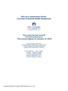
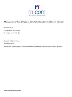
6 This cell-to-cell conduction of depolarization through the myocardium is carried by the fast-moving sodium ions. Repolarization is the return of electrical charges to their original state. This process must happen before the cells can be ready to conduct again. The Conduction System The specialized electrical cells in the heart are arranged in a system of pathways called the conduction system. These specialized electrical cells and structures guide the wave of myocardial depolarization. The conduction system consists of the sinoatrial node (SA node), atrioventricular node (AV node), bundle of His (also called the AV Junction), right and left bundle branches, and Purkinje fibers. Material Protected by Copyright Can Stock Photo Inc. / alila The Sinoatrial (SA) Node The sinoatrial node (also called the SA node or sinus node) is a group of specialized cells located in the posterior wall of the right atrium near the superior vena cava and atrial junction. The SA node normally depolarizes or paces more rapidly than any other part of the conduction system making it the cardiac Pacemaker .
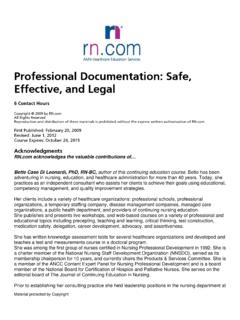
7 It sets off impulses that trigger atrial depolarization and contraction. After the SA node fires, a wave of cardiac cells begin to depolarize. Depolarization occurs throughout both the right and left atria (similar to the ripple effect when a rock is thrown into a pond). This impulse travels through the atria by way of inter-nodal pathways down to the next structure, which is called the AV node. Any malfunction of the sinoatrial node causes a sinus dysrhythmia. The SA node normally fires at a rate of 60-100 beats per minute. The Atrioventricular (AV) Node and AV Junction The next area of conductive tissue along the conduction pathway is at the site of the atrioventricular Material Protected by Copyright (AV) node. This node is a cluster of specialized cells located in the lower portion of the right atrium, above the base of the tricuspid valve. The AV node itself possesses no Pacemaker cells. The AV node has two functions. The first function is to DELAY the electrical impulse in order to allow the atria time to contract and complete the filling of the ventricles.
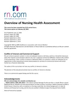
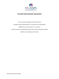
8 The second function is to receive an electrical impulse and conduct it down to the ventricles via the AV junction and bundle of His. Any dysfunction of the AV node results in a junctional or supraventricular dysrhythmia. If the SA node becomes diseased or fails to function properly, the AV node is capable of discharging at an intrinsic rate of 40-60 beats per minute. The AV node normally fires at a rate of 40-60 beats per minute. Can Stock Photo Inc. / alila The Bundle of His After passing through the AV node, the electrical impulse enters the bundle of His (also referred to as the common bundle). The bundle of His is located in the upper portion of the intraventricular septum and connects the AV node with the two bundle branches. The AV node and the bundle of His are referred to collectively as the AV junction. The bundle of His conducts the electrical impulse down to the right and left bundle branches. The bundle branches further divide into Purkinje fibers. Any dysfunction of the Bundle of His results in a bundle branch block dysrhythmia.
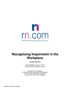
9 Material Protected by Copyright Please note! The Bundle of His is also known as the atrioventricular bundle, and consists of the right and left bundle branches. Can Stock Photo Inc. / alila The Purkinje Fibers At the terminal ends of the bundle branches, smaller fibers distribute the electrical impulses to the muscle cells, which stimulate contraction. This web of fibers is called the Purkinje fibers. The Purkinje fibers penetrate 1/4 to 1/3 of the way into the ventricular muscle mass and then become continuous with the cardiac muscle fibers. The electrical impulse spreads rapidly through the right and left bundle branches and Purkinje fibers to reach the ventricular muscle, causing ventricular contraction, or systole. The Purkinje fibers within the ventricles also have intrinsic Pacemaker ability. Any dysfunction of the Purkinje fibers will result in a ventricular dysrhythmia. Did You Know? The further you travel away from the SA node, the slower the backup pacemakers become.
10 If you only have a heart rate of 30 (from the ventricular back-up Pacemaker ), blood pressure will likely be Material Protected by Copyright low and the patients will likely be quite symptomatic. The Ventricles normally fire at a rate of 20-40 beats per minute. Can Stock Photo Inc. / alila Test Yourself The primary internal Pacemaker of the heart is: A. AV node B. SA node C. Bundle of His Rationale: The sinoatrial node (also called the SA node or sinus node) is a group of specialized cells located in the posterior wall of the right atrium near the superior vena cava and atrial junction. The SA node normally depolarizes or paces more rapidly than any other part of the conduction system making it the cardiac Pacemaker . It sets off impulses that trigger atrial depolarization and contraction. History of Pacemakers Literature written through the 17th and 18th century noted speculation and early experimentation of Material Protected by Copyright electrical impulses in the human body.