Transcription of Common Pacemaker Problems: Lead and Pocket Complications
1 16 Common Pacemaker Problems: Lead and Pocket Complications Enes Elvin Gul and Mehmet Kayrak Selcuk University, Meram School of Medicine, Cardiology Department Konya, Turkey 1. Introduction Pacemakers may cause undesirable Complications during and after implantation. Complications associated with the implantation procedure are uncommon, but include bleeding, infection, or collapsed lung. Generally, each of these problems can be treated quite effectively. Though rare, Pacemaker problems can occur long after the implantation procedure. These "late" Complications include generator failure (extremely rare), and lead failure (less rare). Most of these Complications are uncommon, and can be prevented by simple manoeuvres. Nonetheless, there are some Complications related to Pacemaker system disfunctions which may cause life-threatening Complications . Pacemaker malfunctions may be corrected with accurate, timely diagnosis, but will not be discussed in this chapter.
2 Pacemaker Complications can be divided into acute (immediate) or chronic according to implantation time (or date); lead or Pocket Complications according to the site of complication; and implantation or system failures, according to aethiology (Table 1). The most Common and frequent Complications are those related to implantation. Pacemaker implantation is a safe procedure in experienced hands, but these Complications can be caused even by experienced operators. By following the suggested maintenance schedule, physicians usually detect Pacemaker problems before they become serious. However, it remains important for patients to be aware of the symptoms of bradycardia, symptoms that might indicate a Pacemaker malfunction. Once again, these symptoms include weakness, easy fatigability, lightheadedness, dizziness, and loss of consciousness.
3 Patients experiencing any of these symptoms should notify their doctor. Especially following Complications such as lead dislodgment, pnemothorax, lead indection, and cardiac tamponade, the patients must be informed about these symptoms. The frequency of Complications varies between 10% and 59% for the procedures. This wide range exists due to the Common problem of defining Complications . In some papers, a localized infection or rib fracture is defined as a minor complication. However, in other papers, these Complications are not even recorded. If the Pacemaker lead becomes dislodged on day 4, is this a complication or not? Some authors say yes; others no. This ambiguity leads to a great challenge when trying to compare papers. In this chapter, we will try to discuss these problems systematically and transparently. Modern Pacemakers - Present and Future 300 Significant reductions in frequency of Complications related to pacemakers have been noted due to emerging technological developments, ever increasing experience and patient education.
4 Complications Pocket Complications Lead Complications Pocket hematoma Acute perforation nfection Dislodgement Erosion Infection Migration of Pacemaker Vein thrombosis Twiddler s syndrome Migration Table 1. Classification of Pacemaker Complications . 2. Complications related to the implantation procedure Several lead-related Complications deserve attention, including lead dislodgement, pneumothorax, loose connector pin, conductor coil (lead) fracture, and insulation break. Lead dislodgement Pacing lead displacement and dislodgement is a relatively Common problem and can occur in 5-10 % of the patients (National Pacemaker and ICD database, 2001). Historically, the most Common complication of transvenous pacing has been lead dislodgement. The leads can displace within chambers or out of chambers and should be suspected if the wire appears too taut or too redundant. Leads may dislodge from the initial implant site in the first few days to few weeks following the implantation.
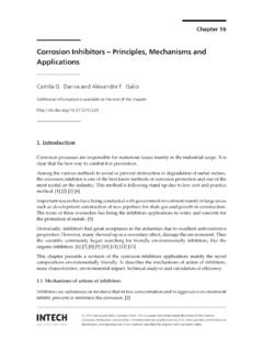
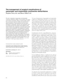
5 Active and passive fixation mechanism of leads help prevent this complication. Atrial lead dislogement is slightly more Common than it is for venrricular leads. Acceptable dislodgmenet rates should be probably be less than 1% for ventricular leads and no more than 2-3% for atrial leads (Braunwald). Although passive fixation leads are stable in the atrial appendage, active fixation leads are necessary to prevent dislodgement in patients with prior cardiac surgery. Lead dislodgement may result in an increase in pacing thresholds, failure to capture, or failure to sense. Lead dislodgement may be radiographically visible or it may be microdislodgement, where there is no radiographic change in position, but there is significant increase in pacing threshold and/or decline in the electrocardiogram amplitude (Figure 1). Also migration of a dislodged lead out of the heart may be associated with thromboembolic Complications if it is not detected acutely.
6 Pacemaker Problems: Lead and Pocket Complications 301 Fig. 1. Top (A): Postero-anterior and lateral chest X-ray films obtained 24 hours after Pacemaker implantation showing atrial lead tip (arrow head) inside the right atrial appendage; Bottom (B): Radiograph obtained three months later showing displacement of atrial lead (arrow head) towards tricuspid annulus. Pneumothorax, hemothorax, and air embolism This complication occurs uncommonly and is directly related to operator experience, the difficulty of the subclavian vein puncture, and is almost eliminated using the cephalic cut-down technique. The incidence of pneumothorax ranges from to % with 80 % of thesecases requiring chest tube placement if > 10 % of the lung is involved, the patient has continued repiratory distress, or hemo-pneumothorax is present (Grier et al., 1990). A pneumothorax estimated to involve < 10 % of the pleural space can probably be observed without chest tube placement.
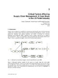
7 This can occur from inadvertent puncture and laceration of the subclavian vein or the subclavian artery or the lung. Modern Pacemakers - Present and Future 302 If a pneumothorax develops, it may manifest during the Pacemaker procedure or as late as 48 hours after implantation (Figure 2). However, these traditional comparisons may become obsolete as the axillary vein cannulation technique (Martin et al., 1996) threatens to eliminate this controversery. To minimize the risk of pneumothorax, fluoroscopic guidance of the subclavian puncture should be used together with careful technique, or to use safe sheats with one-way mechanism which also reduces risk of other potential Complications (hemothorax, inadvertent arterial puncture, air embolism, arteriovenous fstula, thoracic duct injury, and brachial plexus injury). Often pneumothorax is asymptomatic and noted on routine follow-up plain chest radiograph.
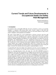
8 Fig. 2. Chest X-ray showing right pneumothorax (arrow) 8 h after placement of a left-sided permanent dual chamber Pacemaker . Location of pneumothorax, ipsilateral or contralateral was reported. Contralateral pneumothorax to the site of the pacing system has been reported, which was secondary to an unsuccessful attempt on that side (Sebastian et al., 2005). In the setting of pneumothorax, pneumopericardium can occur when air dissects through the pulmonary parenchyma along the perivascular sheats back to the hilum. The pericardium is weakest at its reflection surrounding the ostia of the pulmonary veins and air can leak into the pericardial space at this location. In haemodynamically stable patients, CT scan of the chest is investigation of choice (Figure 3). In unstable patients emergency echocardiography may be useful to identify pericardial effusion (Burney et al., 2004). Deep inspiration at the time of central venous access may cause significant air to be drawn into the venous system due to the physiological negative pressure developed.
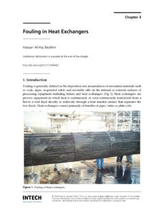
9 Three obligatory conditions need to coexist for pulmonary air embolism to occur: (1) there must be a source of gas/air; (2) an open access to the venous system; and (3) a pressure gradient between the source of gas/air and the venous sytem (Yeakel, 1968). It can be prevented through operator care and using introducers with hemostatic valves. The diagnosis is obvious because it is heralded by a hissing sound as the air is sucked in and with the fluoroscopic confirmation that follows (Figure 4). 100% oxygen should be administered along with ionotropic support in some cases. Aspiration of the embolus from the right heart has also been successful. However, usually no therapy is required, as the air is filtered and consequently absorbed in the lungs. Pacemaker Problems: Lead and Pocket Complications 303 Fig. 3. CT scan of the chest showing massive hemothorax, pneumothorax, and pneumopericardium.
10 Fig. 4. Chest X-ray showing a large air embolus (arrow) in the main pulmonary artery bifurcation during permanent Pacemaker implantation. Modern Pacemakers - Present and Future 304 Three main issues are important in diagnosis and management of this problem : (1) deep inspiration should be avoided in the presence of an open intravenous route; (2) when using a peal-away sheath, temporarily close the intravenous entrance route before inserting the lead; and (3) snoring may be an alarm sound! (Turgeman et al., 2004). Lead perforation Myocardial perforation during lead placement is an uncommon but potentially serious complication. The published incidence of this complication varies from to , but nowadays it is usually lower than 1% (Danik et al., 2007; Carlson at al., 2008). The use of active fixation leads is associated with higher rate of cardiac perforations (Geyfman et al., 2007).