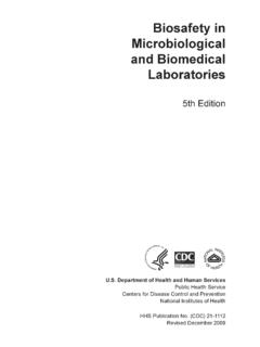
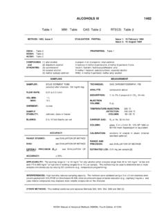
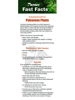
Transcription of Staining thick and thin blood smears
1 Laboratory diagnosis of malaria Staining for malaria parasites Staining thick and thin blood smears Staining blood smears Stain only one set of smears , and leave the duplicates unstained. The latter will prove useful if a problem oc- curs during the Staining process and/or if you wish later to send the smears to a reference laboratory. Giemsa stain - Recommended for detection and identification of blood parasites. 1. Stock 100 Giemsa Buffer M. Na2 HPO4 g NaH2PO4H2O g Deionized water ml Autoclave or filter-sterilize ( m pore). Sterile buffer is stable at room temperature for one year. 2. Working Giemsa Buffer , pH Stock Giemsa Buffer ml Deionized water ml Check pH before use.
2 Should be Stable at room temperature for one month. 3. Triton X-100 5%. Deionized water (warmed to 56 C) ml Triton X-100 ml Prewarm the deionized water and slowly add the Triton X-100, swirling to mix. 4. Stock Giemsa stain (Giemsa stain is available commercially, but the following formulation gives more constant results and does not expire.). Glass beads, mm ml Absolute methanol, acetone-free ml Giemsa stain powder (certified) g Glycerol ml Put into a 500 ml brown bottle the glass beads and the other ingredients, in the order listed. Screw cap tightly. Use glassware that is clean and dry. Place the bottles at an angle on a shaker; shake moderately for 30 to 60 minutes daily, for at least 14.
3 Days. Kept tightly stoppered and free of moisture, stock Giemsa stain is stable at room temperature indefinitely (stock stain improves with age). Just before use, shake the bottle. Filter a small amount of this stock stain through Whatman #1 filter pa- per for use as the working Giemsa stain. 5. Working Giemsa stain ( ): make fresh daily. If a large number of smears are made, stain may need to be changed throughout the day. Working Giemsa buffer 40 ml Giemsa Stain Stock 1 ml 5% Triton X-100 20 l (equivalent to 2 drops). Laboratory diagnosis of malaria Staining thick and thin blood smears Wright (Wright-Giemsa) stain Used in hematology, this stain is not optimal for blood parasites.
4 It can be used if rapid results are needed, but should be followed up when possible with a confirmatory Giemsa stain, so that Sch ffner's dots can be demonstrated. Staining 1. Prepare fresh working Giemsa stain in a Staining jar, according to the previous page. (The 40 ml fills ade- quately a standard Coplin jar; for other size jars, adapt volume but do not change proportions). 2. Pour 40 ml of working Giemsa buffer into a second Staining jar. Add 20 l (2 drops) of Triton X-100. Adapt volume to jar size. 3. Place slides into the working Giemsa stain ( ) for 45-60 minutes. 4. Remove thin smear slides and rinse by dipping 3-4 times in the Giemsa buffer.
5 thick smears should be left in buffer for 5 minutes. 5. Dry the slides upright in a rack. Note: As alternates to this 45-60 minutes in Giemsa stain, the smears could be stained for shorter times in more concentrated stains. One alternate is 10 minutes in 10% Giemsa; the shorter stains yield faster re- sults, but use more stain and might be of less predictable quality. Staining Procedure: Quality Control To ensure that proper Staining results have been achieved, if a positive smear (malaria) is available it may be included with each new batch of working Giemsa stain. Or the specimen being stained may be used as the organisms and/or the white blood cells are a built in quality control.
6 Since good quality control smears are not available commercially, they may be prepared from a patient's blood and stored for future use in the following manner: 1. Choose a patient blood specimen, anticoagulated with EDTA, that has enough parasites so that at least one is found in every two to three fields. 2. Make as many thin smears as possible, preferably within one hour after the blood was drawn from the patient. 3. Allow the smears to dry quickly, using a fan or blower at room temperature. 4. Fix the smears in absolute (100%) methanol; allow them to dry. 5. Wrap in tissue and place them, touching front to back, in a box without separating grooves.
7 6. Label the outside of the box with the species, date and Giemsa control slides.. 7. Store at -70 C (or colder) if the purpose is to make quality control slides or training slides. 8. Just before use, remove the smear from the box and allow the condensation to evaporate; label the slide + malaria and the present date. The smear is now ready for Staining since it was previously fixed.