Transcription of Structure of Human Eye - NCERT
1 St ru ct u re of Hu ma n Eye1 IntroductIonHow many of your family members use spectacles?Let us study how the Human eye uses light and enables us to see objects. Eyes are the most valuable organ of the Human body, which process the images of objects in the vicinity. The eyes interpret size, shape, colour and distance of the objects and give a 3D picture of the objects eyes are seated in a hollow cone shaped cavity (socket) of the Human skull named orbit. The movements of the eye are regulated by six muscles. Fatty tissues in the orbit surrounding, which protect the eyeball, give it flexibility to function.
2 The adnexal structures, like eyebrows, eyelashes, eyelids, protect eyes from foreign elements and liquids maintaining the shape of the eyeballs are called aqueous humour and vitreous eyeball acts like a camera; the image of the objects received by the eyes are conducted to the brain. The visibility by one eye is known as monocular vision, and that by both eyes is binocular you know?Our eyes are sophisticated cameras; they take many pictures in seconds and the brain processes them as messages. Through our eyes, we see the colourful world around us. Unit 117-03-2021 16:54:56 Vision Technician class Xi2 SeSSIon 1: AnAtomy of Human eyeIn this session, you will learn about the parts of Human eye (Fig.)
3 And their functional connections with the Human body. The Human eye is one of the important sensory organs of the Human body. It is very sensitive and exposed to various diseases, thus protection and prevention is necessary to keep the eye safe and healthy. Three layers of Human eye The eyeball has three coats as given fibrous coatThe anterior, transparent, one-sixth part of the eyeball is called cornea. This refracts the rays of light into the eye. Cornea further extends with a membranous Structure called conjunctiva. The connecting area of cornea and conjunctiva is limbus.
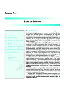
4 external fibrous coat is formed of cornea and vascular coatThis coat is formed by the iris, ciliary body and choroid (anterior to posterior). This coat is vascular and pigmented, underlying the nervous coatInternal nervous coat is formed of retina. The retina receives an inverted image of the objects seen. These images are conducted to the brain through a nerve called the optic nerve, which is connected at the posterior end of the eyeball. Parts of Human eye(a) Anterior chamber: It is the one-third part of the eyeball which is bound by the cornea anteriorly, and the lens posteriorly.
5 It contains the iris and a fluid called the aqueous humour. Fig. : Structure of Eye Unit 230-07-2018 10:37:34 Structure of Human eye3(b) Posterior chamber: It forms the rest of the two-thirds of the eyeball, bound by the intraocular lens anteriorly and optic nerve head and retina posteriorly. It contains a gelly-like fluid called vitreous humour. (c) Pupil: It is an aperture of variable size in the centre of iris, which regulates the amount of light entering the eyeball. (d) Iris: It is the coloured membrane behind the cornea and in point of lens with an aperture of variable size called pupil.
6 It has a circular and long muscle fibre. Iris is attached to the ciliary body. (e) Lens: It is a transparent, biconvex Structure situated between the iris and vitreous humour. Its function is to focus the luminous rays; these rays form a perfect image on the retina. With age, the central portion of the lens compresses by the surrounding fibres and results in opacity, which is called cataract. Blind spot The beginning of the optic nerve in the retina is called the optic nerve head or optic disc. Since there are no photoreceptors (cones and rods) in the optic nerve head, this area of the retina cannot respond to light stimulation.
7 As a result, it is known as the blind spot , and everybody has one in each eye.(f) Vitreous humour: This is a gel-like substance which maintains the shape of the eyeball. It is also a refractive media. (g) Retina: It is a transparent layer forming the inner coat of the eye, it supports the choroid layer. The rays of light, on entering the eyeball, converge and form an image on the fovea the posterior part of the eye on retina. (h) Sclera: It is the outermost coat of the eyeball. It maintains strength and Structure of the eyeball. It is also known as the white of the 330-07-2018 10:37:34 Vision Technician class Xi4(i) Cornea: It is the clear, transparent, anterior portion of the external coat of the eyeball.
8 The rays of light enter this layer. Cornea accounts for two-thirds of the total optical power of the : Parts of Human Eye A. Fill in the blanks1. The _____ is the transparent front part of the The _____ is a gel-like substance that helps to keep the eyeball in its proper The _____is also known as the white of the an eye care unit or clinic with your friends and teacher to observe various charts displaying the anatomy of Your Progress B. Label the parts of the eye in Figure and also list them in the Table given below. SeSSIon 2: fIeld of VISIon And dynAmIc rAnge of Human eyeIn this session, you will learn about the field of vision and the dynamic range of the Human eye.
9 The eyeball acts as a camera and the message of image formation is received and directed to the of vision The field of vision is the area that is seen all around. The field of view of a Human eye is 95 on the left or right of 12345678910 Unit 430-07-2018 10:37:34 Structure of Human eye5the eye, 75 downwards, 60 towards the nose, and 60 upwards ( ). It is in this space that an object can be seen while the eye fixes upon one : Field of vision for Human eyeDid you know?Why do we have two eyes for vision and not just one?There are several advantages of having two eyes instead of one.
10 It gives a wider field of view. A Human being has a horizontal field of view of about 1500 with one eye, and of about 1800 with both eyes. The ability to detect faint objects is, of course, enhanced with two detectors instead of one. Some animals, usually prey animals, have both their eyes positioned on opposite sides of their heads to give the widest possible field of view. But our eyes are positioned on the front of our heads, and it thus reduces our field of view in favour of what is called stereopsis. Shut one eye and the world looks flat-two-dimensional. Keep both eyes open and the world takes on the third dimension of depth.