Transcription of Tracheostomy Decannulation - Respiratory Care
1 Tracheostomy DecannulationHeidi H O Connor MD and Alexander C White MDIntroductionProcess of weaning and Routine DecannulationManaging Accidental DecannulationPost- Decannulation MonitoringDecannulation Failure and Alternatives to DecannulationSummaryTracheostomy tubes are placed for a variety of reasons, including failure to wean from mechanicalventilation, inability to protect the airway due to impaired mental status, inability to manageexcessive secretions, and upper-airway obstruction. A Tracheostomy tube is required in approxi-mately 10% of patients receiving mechanical ventilation and allows the patient to move to astep-down unit or long-term care hospital. The presence of a Tracheostomy tube in the trachea cancause complications, including tracheal stenosis, bleeding, infection, aspiration pneumonia, andfistula formation from the trachea to either the esophagus or the innominate artery. Final removalof the Tracheostomy tube is an important step in the recovery from chronic critical illness and canusually be done once the indication for the tube placement has words: mechanicalventilation; Tracheostomy ; Decannulation ; speaking valve; noninvasive ventilation; long-term care hos-pital.
2 [Respir Care 2010;55(8):1076 1081. 2010 Daedalus Enterprises]IntroductionThe indications for placement of a Tracheostomy tubeinclude failure to wean from mechanical ventilation, im-paired neurologic status, an inability to handle excessivesecretions, and the need to bypass an upper-airway describes the process of trache-ostomy tube removal once the need for the tube has re-solved. There are many advantages to Decannulation ,including improved vocal cord and swallowing patients home or to another care facility is aneasier process if the patient or their caregivers do not needto learn how to manage a Tracheostomy tube. In addition, Decannulation improves patient comfort and perceivedphysical appearance. Patients may also wish to undergodecannulation as part of end-of-life decision of weaning and Routine DecannulationThe placement of a Tracheostomy tube facilitates thetransfer of the patient from the intensive care unit to aweaning facility such as a step-down unit or a long-termcare weaning facilities a multidisciplinary teammanages medical care, rehabilitation, and weaning the pa-tient from prolonged mechanical ventilation.
3 Chronic co-morbidities and the lack of evidence-based weaning andHeidi H O Connor MD and Alexander C White MD are affiliated withthe Department of Pulmonary and Sleep Medicine, Rose Kalman Re-search Center, New England Sinai Hospital, Stoughton, C White MD presented a version of this paper at the 25th NewHorizons Symposium, Airway Management: Current Practice and Fu-ture Directions, at the 55th International Respiratory Congress of theAmerican Association for Respiratory Care, held December 5 8, 2009, inSan Antonio, O Connor has disclosed no conflicts of interest. Dr White has dis-closed a relationship with Breathe : Heidi H O Connor MD, Department of Pulmonary andSleep Medicine, Rose Kalman Research Center, New England SinaiHospital, 150 York Street, Stoughton MA 02072. E-mail: AUGUST2010 VOL55 NO8decannulation guidelines make it difficult to predict wean-ing outcomes of individual stablepatients undergoing prolonged mechanical ventilation usu-ally begin the weaning process by spending increasingamounts of time on a spontaneous breathing trial via hu-midified Tracheostomy mask.
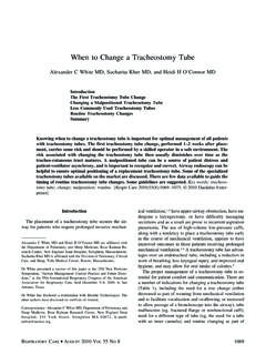
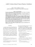
4 Therapist-driven weaningprotocols, such as those involving spontaneous breathingtrials or decreasing levels of pressure support, have beenimplemented in the post-acute-care setting and have beenshown to shorten the time taken to wean patients fromprolonged mechanical ,9 Not all patients are suitable for a weaning protocol, andsome need an individualized approach given the complex-ity of the patient population. The presence of respiratorymuscle weakness, slow recovery from chronic critical ill-ness with multi-organ dysfunction, anxiety from prolongedventilator dependence, chronic anemia, or cardiac dysfunc-tion may necessitate a more gradual weaning strategy. Onemethod involves gradually decreasing the amount of pres-sure-support ventilation prior to withdrawal of full venti-lator support. All patients undergoing weaning from me-chanical ventilation should be carefully monitored usingcontinuous pulse oximetry and cardiac system for patient assessment prior to decannulationis outlined in Figure 1.
5 Once a patient demonstrates theability to tolerate a Tracheostomy mask, it is important toestablish that the upper airway (ie, glottis, vocal cords, andsubglottic space) is patent. The presence of a tracheostomytube can cause complications that may result in upper-airway upper airway can be checkednoninvasively by fully deflating the cuff on the tracheos-tomy tube and placing a gloved finger over the tracheos-tomy tube opening to deflect air through the upper airwayand vocal cords, allowing , tra-cheostomy tube manometry may be used to obtain objec-tive measurements of airway pressures during the use of aspeaking valve or cap. This technique helps identify pa-tients who can tolerate occlusion of the Tracheostomy tubeand also those who may benefit from having a tracheos-tomy tube with a smaller external the patient is unable to phonate, has stridor or laboredbreathing, or manifests any Respiratory distress, a thoroughendoscopic examination of the airway, including the vocalcords and subglottic space, is the air-way patency is compromised by stenosis, granulation tis-sue, or abnormal vocal cord movement, otolaryngologyshould be consulted for further evaluation and initial Tracheostomy tube placed can be up to 8 mminner diameter to facilitate fiberoptic bronchoscopy.
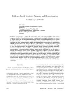
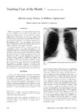
6 If nopathology is found on endoscopy, the tube may be down-sized and changed to a tight-to-shaft (fully deflated) cuffto enhance air flow around the occluded ventilation impairs communication betweenthe patient and caregivers and family, and reduces qualityof ,13In particular, a Tracheostomy tube diverts airflow away from the vocal cords and results in aphonia. Asubstantial percentage of patients recovering from chroniccritical illness also suffer from depression, which mayinfluence their perception of mechanical ventilation andtreatment a patient to regain the func-tion of speech can significantly improve quality of life inpatients who require prolonged mechanical ventilation. Ina spontaneously breathing patient who tolerates occlusionof the Tracheostomy tube as described above, a one-wayspeaking valve may be placed onto the Tracheostomy tubewith a fully deflated cuff. This allows for air flow into thetube during inspiration; however, air flow now exits throughthe upper airway and vocal cords upon exhalation, pro-ducing speech (Fig.)
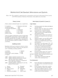
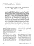
7 2). For those patients who still requirefull mechanical ventilation and are unable to wean, eitheran in-line speaking valve or volume-compensationspeech may be used to allow inspection of the airway, although not es-sential prior to Decannulation , can be helpful. In one study,67% of patients with tracheostomies were found to haveairway abnormalities during airway endoscopy. Findingsincluded tracheal granulomas, tracheomalacia, tracheoste-nosis, and vocal cord of the air-way with the Tracheostomy tube removed allows for visu-alization of the subglottic space and isperformed by retroflexing a fiberoptic rhinolaryngoscopeinto the subglottic space. Some of the abnormalities visu-alized, such as minor mucosal trauma from the tracheos-tomy tube or suction catheter, may not be clinically im-portant and usually do not prevent Decannulation . It hasbeen demonstrated that patients who successfully pass aFig. 1. Decannulation AUGUST2010 VOL55 NO81077tracheostomy-tube-occlusion protocol can be safely de-cannulated without first undergoing fiberoptic is our current practice to routinely inspect thestoma, trachea, subglottic space, and vocal cords either atthe time of a Tracheostomy tube change or prior to decan-nulation (Fig.
8 3). The procedure is safe, requires only top-ical anesthesia, and in a substantial number of patientsidentifies pathology that warrants further otolaryngologyor thoracic surgery input prior to safe Accidental DecannulationAccidental Tracheostomy Decannulation is well describedin the pediatric population, but less well recognized in theadult population. It may be uneventful if the tracheostomytube has been in for some time, the tract has matured, andthe tube can be easily replaced. However, accidental de-cannulation may result in serious consequences if the tra-cheostomy tube was recently placed or if the patient has a difficult ,19We have explored factors associ-ated with accidental Decannulation and have found it maybe related to altered mental status, increased secretions,patient turning, lack of clinically indicated restraints, or apoorly secured Tracheostomy tube (Table 1) (unpublisheddata). Education of staff regarding risk factors for acci-dental Decannulation along with targeted high-risk-patientmonitoring can be effective tools in decreasing the acci-dental Decannulation rate in a long-term care hospital.
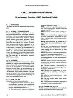
9 Iden-tification of patients with difficult airways, having a sparetracheostomy tube at the bedside, and use of an algorithmfor orotracheal intubation may reduce the morbidity andmortality associated with accidental Decannulation (Fig. 4).The length of time a Tracheostomy tube has been inplace is an important variable in deciding how to bestproceed after an accidental Decannulation occurs. The tra-cheocutaneous tract of a fresh Tracheostomy tube takesup to 7 days to fully form. If during tube reinsertion thecaudal turn is done prematurely, the tube may be inad-vertently placed into the anterior mediastinal space, by-passing the airway. This is more likely to occur if thechange is being performed within 7 days of with an increased neck circumference or shortneck are higher risk of this occurring. Therefore, when anaccidental Decannulation occurs within 7 days of place-ment, translaryngeal intubation may be needed to securethe airway. In some settings, a skilled provider may beable to rapidly reinsert the Tracheostomy tube into the air-way with fiberoptic guidance and avoid the need for trans-laryngeal intubation.
10 Tracheostomy tubes that have beenin place for more than 7 days can usually be easily rein-serted and placement confirmed with fiberoptic inspectionof the 1. Conditions Associated With Accidental TracheostomyDecannulationAltered mental statusIncreased pulmonary secretionsPatient changing position in bedLack of clinically indicated limb restraintsInadequately secured Tracheostomy tubeFig. 2. Biased closed position speaking valve. A: Upon inspirationthe valve opens and air passes through the speaking valve, downthe Tracheostomy tube and into the lungs. B: At the end of inspi-ration the speaking valve returns to a closed position and all ex-haled air is redirected through the upper airway, passing throughthe vocal cords and allowing the patient to speak. (Courtesy ofPassy-Muir.)Fig. 3. Fiberoptic inspection of the trachea (A) and the subglotticspace and vocal cords (B) prior to AUGUST2010 VOL55 NO8 Post- Decannulation MonitoringPrior to a patient undergoing Decannulation it is impor-tant to assess a number of factors to ensure include level of consciousness, cough effectiveness,volume of pulmonary and oral secretions, oxygen require-ments, swallowing function, and ability to tolerate trache-ostomy tube occlusion (Table 2).