Transcription of Treatment of Chondral Defects in the …
1 Of Chondral Defects in the patellofemoral JointAndreas H. Gomoll, MDTom Minas, MD, MSJack Farr, MDBrian J. Cole, MD, MBADrs Gomoll and Minas are from the Cartilage Repair Center, De-partment of Orthopedic Surgery, Brigham and Women s Hospital, Bos-ton, Mass; Dr Farr is from the Cartilage Restoration Center of Indiana, OrthoIndy Knee Care Institute, and Department of Orthopedic Surgery, Indiana University School of Medicine, Indianapolis, Ind; and Dr Cole is from the Cartilage Restoration Center at Rush, Department of Ortho-pedic Surgery, Rush University Medical Center, Chicago, requests: Brian J. Cole, MD, MBA, Rush University Medical Center, 1725 W Harrison, Ste 1063, Chicago, IL 60612. INTRODUCTIONA nterior knee pain is a common musculoskeletal complaint seen daily in the practices of primary care physicians, rheumatologists, and orthopedic surgeons.
2 In the past, the term chondromalacia was misused inter-changeably with anterior knee pain or patellofemoral pain syndrome. The implication that cartilage is the source of symptoms is incorrect as the majority of patients present-ing with anterior knee pain do not have cartilage Defects and cartilage is aneural. The prevalence of patellofemoral cartilage Defects is controversial, as it is unknown what percentage of lesions become symptomatic enough to prompt evaluation. Several studies have reported the pres-ence of high-grade focal Chondral Defects in 11%-20% of knee arthroscopies. Among these Defects , 11%-23% were located in the patella and 6%-15% in the ,11,18 A group investigating asymptomatic NBA basketball play-ers with knee magnetic resonance imaging (MRI) found articular cartilage lesions in 47%, with patellar lesions in 35% and trochlear lesions in 25% of players; however, only approximately half of these Defects were character-ized as high-grade These reports emphasize the importance of a thorough history and physical evalua-tion of the entire kinetic chain from pelvis to foot, a gait analysis, and assessment of all knee structures (tendons, ligaments, and soft tissues) before attributing a patient s symptoms solely to the presence of a Chondral defect.
3 patellofemoral pain, as a subset of anterior knee pain, is typically multifactorial and to achieve success in treat-ment, each contributing factor requires management indi-vidually and in conjunction with the other key to successful Treatment in this group of patients lies not only in the correct diagnosis of a Chondral defect, but more importantly, in the accurate identifi cation of asso-ciated pathomechanical factors, such as patella alta, troch-lea dysplasia, increased lateral position of the tibial tubercle to the femoral sulcus (previously assessed as a Q angle), and secondary soft-tissue problems, such as a weakened or hypoplastic vastus medialis muscle with a contracted lat-eral retinaculum. These pathomechanics lead to abnormal forces of the patellofemoral joint, which can cause injury to the articular cartilage in itself through repetitive micro-trauma or exacerbate the effects of a traumatic comprehensive discussion of anterior knee pain and patellofemoral pain is beyond the scope of this ar-ticle.
4 Therefore, the focus will be on the distinct subset of patients presenting with patellofemoral symptoms who have Chondral Defects . The etiology of these Defects is typically multifactorial, but might include focal degen-eration, trauma (direct impact or repeated patellofemoral instability), and/or repetitive microtrauma with signifi cant biomechanical articular Defects frequently present as anterior knee pain; patients often report their pain to be lo-cated retropatellar, peripatellar, or in the instance of troch-lear Defects , the pain at times is located posteriorly in the popliteal area. As the articular cartilage does not have a nerve supply, the pain reported is always secondary. This secondary pain may be from synovial or capsular irrita-286 THE JOURNAL OF KNEE SURGERYO ctober 2006 / Vol 19 No 4tion or due to subchondral bone overload.
5 Thus, in light of this secondary nature of pain, other factors may also con-tribute, making it diffi cult to assign a percentage of pain to the cartilage pathology. Large Defects can cause click-ing or popping, giving way, and activity-related swelling. Standard patellofemoral symptoms are often reported such as increased pain with prolonged fl exed knee posi-tion and stair climbing. Patients are approximately evenly split in reporting a traumatic versus a more gradual onset of symptoms; sports participation was the most common inciting event associated with the diagnosis of Chondral Patellar dislocation is associated with damage to the articular surface, with Chondral Defects of the patella seen in up to 95% of Patients often report ex-tended courses of physical therapy, bracing and taping, or prior knee ExaminationGait abnormalities, such as intoeing or hip abductor weakness, are frequently seen in this patient population, as is an increase in femoral anteversion and valgus ma-lalignment of the lower extremity.
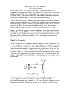
6 Adaptations in gait are also seen such as hip and knee external rotation and contractures of the hip abductors and iliotibial band. Tra-ditionally, the quadriceps angle (Q-angle) has been used in the evaluation of patellofemoral symptoms (Figure 1). Many different methods of measuring this angle have been reported, and the high interobserver variability makes it of questionable ,15 If used, the Q-angle should be evaluated in both full extension and approximately 30 of fl exion, because in some cases, a laterally subluxated patella in full extension can falsely decrease the Q-angle (the patella should be repositioned in the central sulcus before measuring the Q-angle). Quadriceps wasting, es-pecially of the vastus medialis, is common in long-stand-ing patellofemoral symptoms.
7 Recently, more emphasis has been placed on core muscle weakness, especially of the hip abductors, hip extensors, and pelvic stabilizers. Weakness in this group can be demonstrated by asking the patient to single-leg stand on the affected limb, resulting in a pelvic drop on the contralateral side. In addition to poor pelvic support, dynamic internal rotation of the femur and dynamic valgus positioning of the limb can be observed. Activity-related swelling and, in particular, a joint effusion indicate more advanced disease. Palpation of the medial and lateral retinaculum can elicit pain; the lateral structures often are contracted (tested by attempting to reverse patellar tilt), while the medial soft tissues can be attenuated (such as chronic patholaxity of the medial patellofemoral ligament [MPFL]).
8 Patellar mobility, tilt, and subluxation should be assessed and quantifi ed medially and laterally. Catching with mobilization of the patella against the trochlea is sug-gestive of larger Defects . Knee range of motion usually is preserved but may be inhibited by pain or large effusions in acute cases. The J-sign (the patient slowly extends the knee from full fl exion, the patella subluxes laterally once it leaves the constraints of the trochlear groove near full extension) is a common fi nding in normal patients, but if exaggerated it may suggest patholaxity of the medial soft tissues (especially the MPFL).Figure 1. Q-angle: the angle between the lines connecting the center of the patella to the anterosuperior iliac spine proxi-mally and to the tibial tubercle distally.
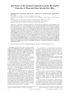
9 Average Q-angles in asymptomatic patients are 14 in males and 17 in Figure 2. Preoperative radiographs in AP (A), lateral (B), and Merchant (C) Defects in the patellofemoral imaging routinely begins by obtaining conven-tional radiographs as a screening tool: standing anteropos-terior (AP), 45 fl exion posteroanterior (PA Rosenberg ), fl exion lateral, shallow angle axial (Merchant), and long-leg axial alignment radiographs (Figure 2). These allow assessment of degenerative changes in the tibiofemoral and patellofemoral articulations, trochlear dysplasia, pa-tella tilt, and subluxation. It is important to bear in mind that although the standard Merchant view is useful to determine joint space narrowing or osteoarthritis of the patellofemoral articulation, it is not effective for the as-sessment of maltracking or trochlear dysplasia.
10 This view is taken at 45 of fl exion where the patella is normally well engaged in the trochlea, whereas maltracking usually occurs from entering the sulcus to 30 of fl exion. Dejour et al13 have shown the advantages of a true lateral radio-graph in assessing trochlear dysplasia and patellar tilt not appreciated on the Merchant accurately assess patella subluxation, computed tomography (CT) of the patellofemoral joint is performed with the leg in full extension, once with the quadriceps relaxed and again with the muscle maximally contracted. Computed tomography also allows a more precise evalu-ation of patellar and trochlear anatomy than the Merchant view. Furthermore, superposition of two CT images, one through the patellofemoral articulation, the other through the tibial tubercle, allows calculation of the tibial tubercle to trochlear groove (TT-TG) distance: the center of the trochlear groove and the center of the tibial tubercle are marked, and the medial-to-lateral distance between the two is measured (Figure 3).