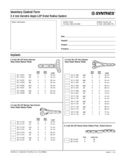
Transcription of Variable Angle LCP Two-Column Volar Distal Radius Plate 2 ...
1 Variable Angle LCP Two-Column Volar Distal Radius Plate For fragment-specific fracture fixation with Variable Angle locking technology. Surgical Technique This publication is not intended for distribution in the USA. Instruments and implants approved by the AO Foundation. Image intensi er control This description alone does not provide sufficient background for direct use of DePuy Synthes products. Instruction by a surgeon experienced in handling these products is highly recommended. Processing, Reprocessing, Care and Maintenance For general guidelines, function control and dismantling of m ulti-part instruments, as well as processing guidelines for i mplants, please contact your local sales representative or refer to: For general information about reprocessing, care and maintenance of Synthes reusable devices, instrument trays and cases, as well as processing of Synthes non-sterile implants, please consult the Important Information leaflet (SE_023827) or refer to: Table of Contents Introduction VA-LCP Two-Column Distal Radius Plate 2.
2 AO Principles 4. Intended Use and Indications 5. Clinical Cases 6. Surgical Technique Three Column Theory 7. Screw Insertion Techniques 8. Screw Angle Overview 10. Approach 12. Implantation 13. Postoperative Treatment/Implant Removal 28. Recommendation 29. Product Information Plates 30. Screws 31. Optional: Trial Implants 33. Instruments 34. Bibliography 38. MRI Information 40. Variable Angle LCP Two-Column Volar Distal Radius Plate Surgical Technique DePuy Synthes 1. Variable Angle LCP Two-Column Volar Distal Radius Plate For fragment-specific fracture fixation with Variable Angle locking technology.
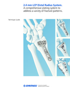
3 Features and Benefits The Variable Angle LCP Two-Column Volar Distal Radius Plate , featuring Variable Angle locking technology, is indi- cated for intra- and extra-articular fractures and osteotomies of the Distal Radius . All implants are available in stainless steel and titanium. Dedicated screws For xation of radial styloid (blue) and support of lunate facet and DRUJ. (green). Bendable Two columns allow independent ne contouring of the radial and intermedi- ate columns Oblong combi-hole Allows accurate Plate positioning on the bone 2 DePuy Synthes Variable Angle LCP Two-Column Volar Distal Radius Plate Surgical Technique Anatomical fit Close to the Volar ridge with rounded Plate edges.
4 Polished surface and countersunk screws to reduce the risk of soft tissue irritation Variable Angle locking Holes allow up to 15 off-axis screw angulation in all directions in order to address the individual fracture patterns Kirschner wire holes Enable preliminary Plate xation Guiding block Allows guided drilling and screw inser- tion in the prede ned nominal Angle Variable Angle LCP Two-Column Volar Distal Radius Plate Surgical Technique DePuy Synthes 3. AO Principles AO PRINCIPLES. In 1958, the AO formulated four basic principles, which have In 1958, the guidelines become AO formulated four basic for internal principles, fixation1,2.
5 Which have become the guidelines for internal xation1, 2. 1 12:08. Anatomic Anatomic reduction fixation Stable fixation Fracture reduction Fracture reduction and and fixation xation to Fracture Fracture fixation xationproviding providingabsolute abso- or to restore restore anatomical anatomical relationships. relationships. relative stability,stability, lute or relative as required as by the prequired atient, theby injury, and thethe the patient, personality injury, 1 2 of andthethe fracture. personality of the fracture. Early, Early, active mobilization mobilization Preservationof Preservation of blood blood supply supply Early and Early and safe mobilization mobilizationand and 4 3 Preservation Preservationof ofthe theblood bloodsupply supply rehabilitation the injured partand rehabilitation of the injured part to to soft tissues and bone bygentle soft tissues and bone by the patient and as a whole.
6 The patient as a whole. reduction techniques gentle reduction and careful techniques and handling. careful handling. 1. M ller ME, M Allg wer, R Schneider, H Willenegger. Manual of Internal Fixation. 3rd ed. Berlin Heidelberg New York: Springer. 1991. 2. R edi TP, RE Buckley, CG Moran. AO Principles of Fracture Management. 2nd ed. Stuttgart, New York: Thieme. 2007. 1. M ller ME, M Allg wer, R Schneider, H Willenegger. Manual of Internal Fixation. 3rd ed. Berlin, Heidelberg, New York: Springer. 1991. 2. R edi TP, RE Buckley, CG Moran. AO Principles of Fracture Management. 2nd ed. Stuttgart, New York: Thieme.
7 2007. 4 DePuy Synthes Expert Lateral Femoral Nail Surgical Technique 4 DePuy Synthes Variable Angle LCP Two-Column Volar Distal Radius Plate Surgical Technique Intended Use and Indications Intended Use The Plate and screw implants included in the Radius Plate product family are intended for temporary fixation, correc- tion or stabilization in the Radius anatomical region. Indications Variable Angle LCP Two-Column Volar Distal Radius Plates are indicated for the fixation of intra- and extra-articular fractures and osteotomies of the Distal Radius . Variable Angle LCP Two-Column Volar Distal Radius Plate Surgical Technique DePuy Synthes 5.
8 Clinical Cases Case 1. 24-year-old male with AO fracture, fall from scaffold Preoperative, AP view Preoperative, lateral view Postoperative, AP view Postoperative, lateral view Case 2. 77-year-old female with AO 23C1 fracture, fall Preoperative, AP view Preoperative, lateral view Postoperative, AP view Postoperative, lateral view, 20 inclined 6 DePuy Synthes Variable Angle LCP Two-Column Volar Distal Radius Plate Surgical Technique Three Column Theory The treatment of Distal Radius fractures requires a meticulous reconstruction of the joint surface, as well as stable internal fixation and early functional post-operative treatment.
9 Extra-articular fractures require both the restoration of the Volar tilt and radial length to reduce the possibility of dis- placement. Any malalignment may result in limitations of movement, changes of load distribution, mid-carpal instabil- ity as well as the increased risk of osteoarthritis in the radio- carpal joint. Intra-articular fractures with articular displacement of more than 2 mm in the radiocarpal joint inevitably result in osteo- arthritis and functional impairment. The Distal Radius and Distal ulna form a three-column biome- chanical construction 3: The ulnar column is the Distal ulna, the triangular fibrocar- tilage and the Distal radio-ulnar joint.
10 The intermediate column is the medial part of the Distal Radius , with the lunate fossa and the sigmoid notch. The radial column is the lateral Radius with the scaphoid VA-LCP Two-Column Volar Distal Radius Plate allows both fixation and fossa and the styloid process. buttressing of the two columns of the Distal Radius A dorsally displaced fracture of the Distal Radius indicates not only dorsiflection in the sagittal plane, but also radial devia- tion in the frontal plane and supination in the transverse radial column plane. Intermediate column Following reduction, stabilization requires optimal fixation of the intermediate column as well as the radial column.