Transcription of Western Blotting Transfer and Detection Procedure
1 PROTOCOL 1850 Millrace Drive, Suite 3A Eugene, Oregon 97403 Telephone: 1-800-910-6486 or 541-284-1800 Fax: 541-284-1801 Customer Service: Tech Support: Western Blotting Transfer and Detection Procedure 10-10 DESCRIPTION Western Blotting Transfer and Detection Procedure ADDITIONAL MATERIALS REQUIRED Reagents: MitoSciences primary antibody(ies) Secondary antibody Phosphate buffered saline, PBS (recipe see Page 6) Blocking buffer : 5% fat free milk in PBS PVDF membrane m Immobilon-P (Millipore IPVH00010) Tween-20 (Aldrich 27,434-8) CAPS (Sigma C2632) Methanol Double distilled water Equipment: Vertical acrylamide electrophoresis unit (BioRad mini Protean series recommended) Electroblotting unit-fully submerged (BioRad mini Protean series recommended) pH meter Weighing balance Other standard lab equipment SAMPLE PREPARATION 1.
2 For performing Western Blotting Transfer and Detection , it is always recommended to isolate mitochondria from cells before analysis. Protocols for mitochondrial isolation can be found at It is possible to probe whole tissue or cell extract, but this may result in a weaker signal and/or additional bands resulting from non-specific cross-reactivity to non-mitochondrial proteins. In the experience of MitoScience scientists, such non-specific cross-reactivity is limited. 2. Samples should be solubilized in the SDS-PAGE loading buffer (preparation on Page 6). 3. After solubilization the sample should be spun at 13,000 rpm for 5 minutes. 4. The supernatant should be collected and loaded onto the gel. Note: In most cases the sample need not be heated/boiled in this sample buffer prior to loading. However sample heating may be incorporated into this protocol if desired, see Page 8.
3 MitoSciences antibodies have been optimized for the Detection of mitochondrial proteins over a wide range of antigen concentrations; however suggested concentrations are presented in Western Blotting Protocol Telephone: 1-800-910-6486 or 541-284-1800 Fax: 541-284-1801 Customer Service: Tech Support: Page | 2 of 9 Table 1 providing the user with the opportunity of producing an optimal signal. Using these suggested sample amounts should yield a linearly proportional signal that allows for quantitation. Sample Loading Heart mitochondria 1-10 g/lane Muscle mitochondria 1-10 g/lane Brain mitochondria 5-20 g/lane Other tissue mitochondria 5-20 g/lane Muscle tissue extract 5-20 g/lane Non-muscle tissue extract 10-30 g/lane Cultured cell mitochondria 10-30 g/lane Cultured cell extract 10-30 g/lane Table 1.
4 Suggested protein loading concentrations. Using these loading amounts as simple guidelines in conjunction with the recommended electrophoresis, Transfer , and immunodetection methods detailed in this manual will yield optimal, reproducible, and quantitative Western Blotting results with MitoSciences monoclonal antibodies. ACRYLAMIDE GEL PREPARATION AND ELECTROPHORESIS COMMERCIALLY AVAILABLE GELS Most commercially available pre-cast gel systems are suitable for use with this protocol. As an example excellent pre-cast Tris-glycine gels in acrylamide single step or gradient are available from Invitrogen and Biorad (examples EC61355 BOX and 161-1124, respectively). These Tris-glycine gels can be used with the electrophoresis buffers detailed in this guide. Other buffered gel systems may require specific electrophoresis buffers.
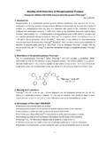
5 Electrophoresis using pre-cast systems such as these should always be performed according to the manufacturer s instructions. PURING GELS BY HAND Alternatively, if desired acrylamide gels can be poured by hand. While it is possible to use a single acrylamide concentration such as a straight 15% gel, MitoSciences highly recommends the use of a linear acrylamide concentration such as 10-20% which will give optimal resolution of all proteins between 10 kDa and 100 kDa. A recipe for pouring 10-20% acrylamide gels in the a 10x gel BioRad Mini-PROTEAN II multicasting chamber is detailed in Table 2 when using a two chamber gradient mixer. BioRad Mini Protean II gel Typical gradient former casting chamber (165-2950) Western Blotting Protocol Telephone: 1-800-910-6486 or 541-284-1800 Fax: 541-284-1801 Customer Service: Tech Support: Page | 3 of 9 10% acrylamide 20% acrylamide mL 30% acrylamide 21 mL 30% acrylamide 12 mL ddH20 1 mL ddH20 180 L 10% SDS 180 l 10% SDS 9 mL M Tris pH 9 mL M Tris pH 180 L 10% APS 180 L l 10% APS 18 L TEMED 18 L TEMED Total volume 32 mL Total volume mL Table 2.
6 Recommended acrylamide BioRad 30% Acrylamide/Bis Solution :1 (161-0158). Once poured, cover the gels in 50% isopropanol solution. Once all 10 gels have set, pour off the isopropanol, rinse with water and remove gels from casting chamber. Now a stacking gel and comb are used. pH Stacking gel mL 30% acrylamide mL ddH20 28 L 10% SDS mL 1 M Tris pH 28 L 10% APS 5 L TEMED Total volume 5 mL Table 3. Stacking gel Samples should be loaded into wells and it is highly recommended to load at least one well with prestained molecular weight standards such as Invitrogen Multimark (LC5725) or BioRad Kaleidoscope (161-0375) markers. Electrophoresis conditions vary, however the samples should be separated at 150V for approximately 2 hours or until the sample buffer, bromophenol blue dye, has almost run off the bottom of the gel.
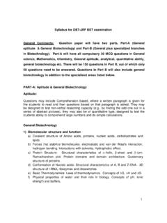
7 A recipe for electrophoresis running buffer is described in Page 6. ELECTROBLOTTING Electroblotting should be performed with a fully submerged system such as BioRad Mini Trans-blot system*. Additionally, MitoSciences highly recommends using the CAPS buffer Transfer system**. The recipes for all buffers are detailed in Page 6. Also highly recommended is the use of a PVDF membrane such as Immobilon rather than a nitrocellulose membrane. After electrophoresis is finished the gel should be soaked in CAPS Transfer buffer for 30 minutes before assembling the Transfer sandwich detailed in Figure 1. Western Blotting Protocol Telephone: 1-800-910-6486 or 541-284-1800 Fax: 541-284-1801 Customer Service: Tech Support: Page | 4 of 9 Electroblotting should be carried out at 150 mAmp for 2 hours.
8 Good electrophoretic Transfer is indicated by the complete Transfer of prestained molecular weight markers below 100 kDa. Prestained markers of greater than 100 kDa may not Transfer completely from the gel onto the membrane. It is also possible to use a semi-dry system such as Hoefer TE-70 however results are not guaranteed. The possibility also exists to use a Tris-glycine or Towbin buffer for electroblotting however results with individual antibodies may vary from the established CAPS Transfer method. BLOCKING AND IMMUNODETECTION 1. Membranes should be blocked for at least 3 hours in 5% milk/PBS solution, although blocking overnight at 4oC is recommended. 2. The membrane should be washed for 10 minutes in PBS Tween-20. 3. The membrane can now be incubated with the primary MitoSciences mouse monoclonal antibody(ies).
9 Antibodies should be diluted to the recommended concentration in a 1% milk/PBS incubation solution (Note: Except for MitoSciences ANT mAb, MSA02, which is provided with its own incubation solution). An antibody solution of 5 mL should be enough to cover a 50 cm2 membrane in a sealed bag or tube with constant rocking/agitation/rolling for 2 hours is recommended. 4. Now wash the membrane in PBS Tween-20 solution for 5 minutes. 5. Repeat the above step twice more. Figure 1. Assembly of Electroblotting sandwich in submerged Transfer apparatus. Western Blotting Protocol Telephone: 1-800-910-6486 or 541-284-1800 Fax: 541-284-1801 Customer Service: Tech Support: Page | 5 of 9 6. Now incubate the membrane with the secondary antibody, which should be an anti-mouse antibody, for 2 hours. Note: This antibody should also be conjugated appropriately for the Detection method of choice.
10 Two highly recommended methods are alkaline phosphatase (AP) and horseradish peroxidase conjugated secondary antibodies (see below). Use this antibody at the dilution recommended by the manufacturer in a 1% milk/PBS solution. Inclusion of sodium azide as a preservative in this solution or subsequent solutions will inhibit the activity of horseradish peroxidase conjugated antibodies. 7. Now wash the membrane in PBS Tween-20 solution for 5 minutes. 8. Repeat the above step twice more. Note: The blot must be rinsed in PBS to remove any Tween-20, which can be inhibitory to the Detection method. 9. The blot is now ready for development. Development With An Alkaline Phosphatase Conjugated Secondary Antibody. Membrane should be incubated in AP color development buffer supplemented with 1% v/v BCIP and 1% v/v NBT (all three solutions are supplied by BioRad as product #170-6432).