Transcription of Zimmer MIS Multi-Reference Instrumentation
1 For NexGen Cruciate Retaining & NexGen Legacy Posterior Stabilized KneesZimmer MIS Multi-Reference 4-in-1 Femoral InstrumentationSurgical TechniqueZimmer MIS Multi-Reference 4-in-1 Femoral Instrumentation Surgical Technique1 Table of ContentsAbbreviated Surgical technique 2 Introduction 4 Preoperative Planning 5 Surgical Approach 5 Patient Preparation 6 Incision and Exposure 6 MIS Midvastus Approach 7 MIS Subvastus Approach 8 MIS Medial Parapatellar Arthrotomy 9 Surgical technique 10 Establish Femoral Alignment 10 Cut the Distal Femur 12 Size Femur & Establish External Rotation 13 Finish the Femur 15 Anterior Referencing technique 15 Posterior Referencing technique 17 MIS Notch/Chamfer Trochlear Guide 19 MIS QS Notch Guide 21 Resect Proximal Tibia 22 Check Flexion/Extension Gaps 27 Prepare the Patella 28 Resect the Patella 29 Finish the Patella 31 Patella Protectors 32 Perform a Trial Reduction 33 Tibial Position based on Anatomic Landmarks 34 Perform a Trial Reduction 35 Implant Components 37 Close Incision 41 Appendix 1 42 Surgical technique For MIS Multi-Reference 4-in-1 Femoral InstrumentationZimmer MIS Multi-Reference 4-in-1 Femoral Instrumentation Surgical Technique25aSet External Rotation
2 (Anterior Referencing)1 Drill 8mm Pilot Hole2 Insert and Secure Mini Distal Femoral Cutting Guide5bSet External Rotation (Posterior Referencing) Zimmer MIS Multi-Reference 4-in-1 Femoral Instrumentation Surgical Technique31432 Size the Femur3 Cut Distal Femur4 Set External Rotation (Posterior Referencing)6 Place Femoral Finishing Guide; Adjust M/L & Pin (Anterior Referencing)Finish the Femur1. Anterior condyles2. Posterior condyles3. Posterior chamfer4. Anterior chamfers7 Zimmer MIS Multi-Reference 4-in-1 Femoral Instrumentation Surgical Technique4 IntroductionSuccessful total knee arthroplasty depends in part on re-establishment of normal lower extremity alignment, proper implant design and orientation, secure implant fixation, and adequate soft tissue balancing and stability.
3 The NexGen Complete Knee Solution and Multi-Reference 4-in-1 Instruments are designed to help the surgeon accomplish these goals by combining optimal alignment accuracy with a simple, straight-forward instruments and technique assist the surgeon in restoring the center of the hip, knee, and ankle to lie on a straight line, establishing a neutral mechanical axis. The femoral and tibial components are oriented perpendicular to this axis. Femoral rotation is determined using the posterior condyles or epicondylar axis as a reference . The instruments promote accurate cuts to help ensure secure component fixation.
4 Ample component sizes allow soft tissue balancing with appropriate soft tissue femur, tibia, and patella are prepared independently, and can be cut in any sequence using the principle of measured resection (removing enough bone to allow replacement by the prosthesis). Adjustment cuts may be needed later. The Multi-Reference 4-in-1 instruments provide a choice of either anterior or posterior referencing techniques for making the femoral finishing cuts. The anterior referencing technique uses the anterior cortex to set the A/P position of the femoral component. The posterior condyle cut is variable. The posterior referencing technique uses the posterior condyles to set the A/P position of the femoral component.
5 The variable cut is made Mini-Incision TKA technique has been developed to combine the alignment goals of total knee arthroplasty with less disruption of soft tissue. To accommodate this technique , some of the original Multi-Reference 4-in-1 Instruments have been modified. However, if preferred, a standard incision can be used with the instruments. Prior to using a smaller incision, the surgeon should be familiar with implanting NexGen components through a standard knee arthroplasty using a less invasive technique is suggested for nonobese patients with preoperative flexion greater than 90 . Patients with varus deformities greater than 17 or valgus deformities greater than 13 are typically not candidates for an MIS refer to the package inserts for complete product information, including contraindications, warnings, precautions, and adverse effects.
6 Zimmer MIS Multi-Reference 4-in-1 Femoral Instrumentation Surgical Technique56 Transverse AxisMechanical Axis90 Preoperative PlanningUse the template overlay (available through your Zimmer Representative) to determine the angle between the anatomic axis and the mechanical axis. This angle will be reproduced intraoperatively. This surgical technique helps the surgeon ensure that the distal femur will be cut perpendicular to the mechanical axis and, after soft tissue balancing, will be parallel to the resected surface of the proximal tibia. Surgical ApproachThe femur, tibia, and patella are prepared independently, and can be cut in any sequence using the principle of measured resection (removing enough bone to allow replacement by the prosthesis).
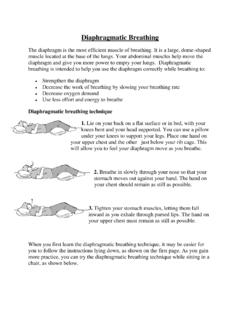
7 Adjustment cuts may be needed MIS Multi-Reference 4-in-1 Femoral Instrumentation Surgical Technique6 Patient PreparationTo prepare the limb for MIS total knee arthroplasty, adequate muscle relaxation is required. This may be accomplished with a short-acting, nondepolarizing muscle relaxant. The anesthesiologist should adjust the medication based on the patient s habitus and weight, and administer to induce adequate muscle paralysis for a minimum of 30-40 minutes. It is imperative that the muscle relaxant be injected prior to inflation of the tourniquet. Alternatively, spinal or epidural anesthesia should produce adequate muscle desired, apply a proximal thigh tourniquet and inflate it with the knee in hyperflexion to maximize that portion of the quadriceps that is below the level of the the patient is draped and prepped on the operating table, determine the landmarks for the surgical incision with the leg in extension.
8 Incision and ExposureThe incision may be made with the leg in extension or flexion depending on surgeon preference. The surgeon can choose a midvastus approach, a subvastus approach, or a parapatellar medial arthrotomy. Also, depending on surgeon preference, the patella can be either everted or length of the incision is dependent on the size of the femoral component needed. Although the goal of a MIS technique is to complete the surgery with an approximately 10cm-14cm incision, it may be necessary to extend the incision if visualization is inadequate or if eversion of the patella is not possible without risk of avulsion at the tibial tubercle.
9 If the incision must be extended, it is advisable to extend it gradually and only to the degree necessary. The advantage of a MIS technique is dependent on maintaining the extensor mechanism a slightly oblique parapatellar skin incision, beginning approximately 2cm proximal and medial to the superior pole of the patella, and extend it approximately 10cm to the level of the superior patellar tendon insertion at the center of the tibial tubercle (Fig. 1). Be careful to avoid disruption of the tendon insertion. This will facilitate access to the vastus medialis obliquis, and allow a minimal split of the muscle.
10 It will also improve visualization of the lateral aspect of the joint obliquely with the patella everted. The length of the incision should be about 50% above and 50% below the joint line. If the length of the incision is not distributed evenly relative to the joint line, it is preferable that the greater portion be 1 Divide the subcutaneous tissue to the level of the retinaculum. NOTE: Using electrocautery to complete the exposure will help minimize bleeding after deflation of the tourniquet, as well as late muscle MIS Multi-Reference 4-in-1 Femoral Instrumentation Surgical Technique7 MIS Midvastus ApproachMake a medial parapatellar incision into the capsule, preserving approximately 1cm of peritenon and capsule medial to the patellar tendon.