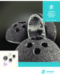
Transcription of Zimmer Osteochondral Allograft,Zimmer …
1 Zimmer Chondrofix Osteochondral Allograft Surgical TechniqueTOCTOCP rocedures for the Treatment of Osteochondral Defects in the Femoral CondyleTable of ContentsIntroduction Arthroscopic or Mini Arthrotomy Procedures for the Treatment of Osteochondral Defects in the Femoral Condyle The Zimmer Chondrofix Osteochondral Allograft is a decellularized allograft consisting of hyaline cartilage and cancellous bone. The grafts are provided precut 10mm in length and 7mm, 9mm, 11mm or 15mm in diameter. Grafts and instrumentation kits are color matched by size7mm Red9mm Blue11mm Purple15mm OrangeSECTION PAGE2 Chondrofix Osteochondral Allograft Surgical Technique #2 5-7 Mini Arthrotomy Repair of Osteochondral Lesions in the Knee Using Cannulated Instrumentation 5-7 SECTION PAGE1 Chondrofix Osteochondral Allograft Surgical Technique #1 1-4 Arthroscopic Repair of Osteochondral Lesions in the Knee Using Punch Instrumentation 1-4 Warnings and Precautions: For use only by surgeons familiar with Osteochondral autograft or allograft implantation procedures.
2 Do not re-sterilize the graft. For single-patient use only. For single-occasion use only. Do not use the graft if damaged or expired. Do not use the graft if sterility is compromised. Do not use the graft if package integrity has been compromised. For use with Zimmer instrumentation only. Instruments kits are single-use. Do not re-use single-use instruments. Instruments are provided sterile. Do not re-sterilize single-use instruments. Do not use an instrument if damaged. Do not use a sterile instrument if past the labeled expiration date. Do not use if instrument sterility or package integrity has been Repair of Osteochondral Lesions Using Punch Instrumentation1 Establish Arthroscopic Portals Establish arthroscopic portals that optimize access to the lesion. To ensure portal placement is perpendicular to lesion, you may want to use an 18 gauge spinal needle as a guide.
3 Consider debriding the fat pad to allow for easier access to the lesion. With the scope in place, examine the affected articular cartilage to be treated (Fig. 1). Fig. 2 Fig. 3 Determine the Size of the Lesion 2 and Necessary Graft ConfigurationDetermine the size of the lesion and necessary graft configuration by covering the defect area with a Sizer(s) of corresponding diameter. The Sizer kit includes instruments of 7mm, 9mm, 11mm and 15mm diameters. Insert the Sizer into the joint and cover the lesion area with the tip of the Sizer (Fig. 2). Once the size of the lesion and graft configuration has been determined, remove the Sizer and retain for Step 1 Select the Appropriate Graft 3 and InstrumentationSelect the appropriate diameter graft(s) and corresponding instrumentation kit(s). Measure the length of the graft(s) from the apex to the bone base to determine maximum depth of the recipient hole (Fig. 3). Caution: Slight variability exists among graft lengths.
4 It is important to measure the lengths of ALL grafts being used prior to creating the recipient site. If implanting multiple overlapping grafts, consider creating a shallower recipient the articular cartilage being the size of the the appropriate size graft(s).SECTION12 Arthroscopic Repair of Osteochondral Lesions Using Punch InstrumentationFig. 4 Fig. 54 Prepare the Recipient SitePrepare the recipient site by introducing the Punch with the Obturator into the portal and locating the lesion site. When positioned at the lesion site, remove the Obturator from the Punch by twisting the Obturator 45 degrees CCW to unlock. This will expose the cutting edge of the Punch to the articular cartilage. Replace the Obturator with the Impaction Cap and lock into place; twist 45 degrees CW (Fig. 4).Caution: Do not mallet the Punch without the Impaction Cap in place. Caution: Before advancing the Punch, make sure to position it perpendicular to the articular cartilage surface at the lesion site.
5 The Punch has circumferential markings at the cutting end in 2mm increments from 2mm 12mm to aid in depth identification and perpendicularity assessment. With the Impaction Cap in place, mallet the top surface of the cap to advance the Punch into the bone. Periodically check the depth measurements around the circumference to ensure perpendicular alignment. Caution: Do not drive the Punch to a depth greater than the length of the longest graft to be used. 5 Remove the Bone Within the PunchOnce the Punch has been advanced to the appropriate depth, remove the Impaction Cap. Connect the Drill Bit of corresponding size to the chuck on the surgical drill. Insert the Drill Bit into the shaft of the Punch. The Punch will function as the drill guide maintaining axial alignment between the Drill Bit and the drill. Advance the Drill Bit into the bone to remove the bone within the Punch until the physical stop on the Drill Bit contacts the Punch handle (Fig.)
6 5). Remove the Punch and Drill Bit assembly. Inspect the recipient site for loose debris; remove if the recipient Drill Bit to remove bone within the Repair of Osteochondral Lesions Using Punch Instrumentation Verify the Depth of 6 the Recipient Site Before implanting the graft, verify the depth of the recipient site with the Sizer of corresponding diameter used in Step 2 (Fig. 6). Chamfer the leading edge of the bone portion of the graft with rongeurs or rasps. If necessary, the bone portion of the graft can be trimmed with rongeurs, small bone saws or bone cutters to match the depth of the recipient site. Fig. 6 Fig. 7 Fig. 8 Loading the Graft into 7the Delivery DeviceWith Delivery Device in hand, depress the Plunger until the middle of the colored band on the Plunger shaft is positioned at the opening of the Delivery Device T-handle. This will expand the metal springs at the open end of the Delivery Device and allow for insertion of the graft.
7 Make sure to insert the cartilage end of the graft first into the open end of the Delivery Device (Fig. 7). Once the graft is inserted, retract the Plunger in order to contract the metal springs to hold the graft. At this point the graft is captured in the Delivery Device and is ready for implantation. 8 Implanting the GraftIntroduce the Delivery Device into the surgical site. When the Delivery Device is positioned over the recipient site, release the graft into the recipient hole by depressing the Plunger on the Delivery Device (Fig. 8). The Delivery Device will leave the graft 1mm proud in the recipient hole. To advance the graft the remaining 1mm, remove the Delivery Device and insert the Tamp of corresponding size (Fig. 9). Gently tap the head of the Tamp until the graft is flush with the surrounding articular cartilage. Caution: When using a Chondrofix Allograft, it is important to make sure the graft is flush with and matches the surface contour of the surrounding articular Tamp.
8 Fig. 9 Using Sizer, verify depth of recipient the cartilage end of the graft into the open end of the Delivery the Plunger on the Delivery Tamp and gently tap until graft is BandSECTION14 Arthroscopic Repair of Osteochondral Lesions Using Punch Instrumentation9 Close the PortalOnce all the grafts have been implanted, do a final check of the graft position relative to the surrounding cartilage. Close the portals and drain the joint through the superior portal. 10 Post-Operative CareThe rehabilitation protocol is at surgeon discretion. General post-operative guidelines include:Multi-Graft Implantation TechniqueRepeat Steps 3- 8. Warning: Graft is not recommended for Mosaicplasty graft constructs without proper shouldering of surrounding host : Prior to reusing a Punch or Drill Bit, inspect the cutting edges of the instruments to ensure that the surfaces have not been BearingROME xercisePhase I 0-6 weeksWeight bearing as toleratedProgress ROM as tolerated, consider CPM Machine (condylar)Biking with no resistance, straight leg raises, open chain exercisesPhase II 6-12 weeksFull weight bearingIncrease ROM, progress to full ROMC losed chain exercises, initiate weight shifts, begin w/b exercises weeks 8-10 progressing to week 12 Phase III12-26 weeksFull weight bearingContinue progressive exercises1 SECTIONA rthroscopic Repair of Osteochondral Lesions Using Punch Instrumentation2 SECTIONMini Arthrotomy Repair of Osteochondral Lesions Using Cannulated Instrumentation 51 Create a Mini Arthrotomy Create a medial or lateral arthrotomy based on lesion 1 Fig.
9 2 Determine the Size of the Lesion 2 and Necessary Graft ConfigurationDetermine the size of the lesion and graft configuration by covering the defect area with a Sizer(s) of corresponding diameter. The Sizer kit includes instruments of 7mm, 9mm, 11mm and 15mm diameter. Insert the Sizer into the joint and cover the lesion area with the tip of the Sizer (Fig. 1). Once the lesion size and graft configuration have been determined, remove the Sizer and retain for Step 6. Fig. 3 Select the Appropriate Graft 3 and InstrumentationSelect the appropriate diameter graft(s) and corresponding instrumentation kit(s). Measure the length of the graft(s) from the apex to the bone base to determine maximum depth of recipient hole (Fig. 2). Caution: Slight variability exists among graft lengths. It is important to measure the lengths of ALL grafts being used prior to creating the recipient site. If implanting multiple overlapping grafts, consider creating a shallower recipient Prepare the Recipient SitePrepare the recipient site.
10 In a mini-arthrotomy procedure, the Sizer and Guide Pin function similarly to the Punch used in the arthroscopic procedure with the Guide Pin determining the depth of the recipient site and the Sizer assisting in perpendicular alignment. Slide the Guide Pin through the hole in the center of the Sizer. Locate the center of the lesion site with the Guide Pin. Orient the Sizer perpendicular to the cartilage : Perpendicular positioning during recipient site creation is critical to this procedure and the appropriate fit of Chondrofix Osteochondral the Guide Pin into the lesion to the desired depth (Fig. 3). Guide Pin depth should be approximately 10mm greater than the recipient site depth in order to ensure stability during reaming. Caution: Do not over advance the Guide size of the the appropriate size graft(s).Advance Guide Pin into the Repair of Osteochondral Lesions Using Punch InstrumentationSECTION2 Mini Arthrotomy Repair of Osteochondral Lesions Using Cannulated Instrumentation 6 Color BandFig.