Fluorescence Microscopy
Found 9 free book(s)
Polymer-Modified Asphalt 101
pavement.engineering.asu.eduNov 12, 2014 · Fluorescence Microscopy Optical reflective fluorescence photomicrograph of a swollen polymer-asphalt blend. Initial Dispersion of Polymer in Asphalt . Fluorescence Microscopy Formation of Polymer Networks in Asphalt . Fluorescence Microscopy Maturing of Polymer Network in Asphalt .
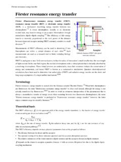
Förster resonance energy transfer
atlas.physics.arizona.eduIn fluorescence microscopy, fluorescence confocal laser scanning microscopy, as well as in molecular biology, FRET is a useful tool to quantify molecular dynamics in biophysics and biochemistry, such as protein-protein interactions, protein–DNA interactions, and protein conformational changes. For monitoring the complex formation
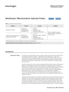
MitoTracker® Mitochondrion-Selective Probes
tools.thermofisher.comanalysis, or fluorescence microscopy. If immobilized cells on coverslips are needed, use poly-D-lysine to coat the slides or coverslips before mounting. If the cells are to be fixed and permeabil ized, continue to Fixation and Permeabilization after Staining. Optional: Fixation and Permea-

EdU (5-ethynyl-2’-deoxyuridine)
tools.thermofisher.comFluorescence microscopy (Imaging) 50 coverslips Alexa Fluor® 488 dye C10337 • Includes blue-fluorescent nuclear counterstain, Hoechst 33342 • Not interchangeable with flow cytometry assays Alexa Fluor® 555 dye C10338 Alexa Fluor® 594 dye C10339 Alexa Fluor® 647 dye C10340 Table 4. Dye- and hapten-containing azides.
Fundamentals of Scanning Electron Microscopy and Energy ...
nanotechftm.tmf.bg.ac.rsNFMC Spring School on Electron Microscopy, April 2011 FWHM2=kE+FWHM noise 2 XEDS: Artifacts Si “escape peak” † 1.74keV bellow true characteristic peak position NFMC Spring School on Electron Microscopy, April 2011 The internal fluorescence peak † A small Si KDpeak at 1.74keV The sum peak † too high count rate
Characteristics of Benign & Malignant Neoplasms
www.columbia.edu-light microscopy: biopsy-cytology (Fine Needle Aspiration—FNA)-immunohistochemistry-fluorescence in situ hybridization (FISH)-molecular probes, incl. gene microarray-flow cytometry (lymphomas, leukemias) 10 Tumor Markers *Molecules in plasma produced by tumor cells
GRAPHENE OXIDE DETECTION IN AQUEOUS SUSPENSION
www.thecompleteguidetohealth.coma) Bright field. b-d) Fluorescence extinction microscopy (FQM) Kim et al, 2010. Seeing graphene-based sheets, Materials Today, Volume 13, 2010, Pages 28-38, Literature Image Low magnification TEM "The figure shows a TEM image of bilayer graphene with edges that tend to curl and bend slightly" 5

Fluorescence Excitation and Emission Fundamentals
www.chem.uci.eduFluorescence was first encountered in optical microscopy during the early part of the twentieth century by several notable scientists, including August Köhler and Carl Reichert, who initially reported that fluorescence was a nuisance in ultraviolet microscopy.

The Handbook - Laboratory Diagnosis of Tuberculosis by ...
www.who.intmicroscopy of two consecutive sputum specimens will identify the vast majority (95–98%) of smear-positive TB patients**. Moreover, microscopy can be decentralised to peripheral laboratories. Despite its advantages sputum smear microscopy does fall short in test sensitivity, especially for certain patient groups such as those living