Transcription of Fluorescence Excitation and Emission Fundamentals
1 Fluorescence Excitation and Emission Fundamentals Fluorescence is a member of the ubiquitous luminescence family of processes in which susceptible molecules emit light from electronically excited states created by either a physical (for example, absorption of light), mechanical (friction), or chemical mechanism. Generation of luminescence through Excitation of a molecule by ultraviolet or visible light photons is a phenomenon termed photoluminescence, which is formally divided into two categories, Fluorescence and phosphorescence, depending upon the electronic configuration of the excited state and the Emission pathway.
2 Fluorescence is the property of some atoms and molecules to absorb light at a particular wavelength and to subsequently emit light of longer wavelength after a brief interval, termed the Fluorescence lifetime. The process of phosphorescence occurs in a manner similar to Fluorescence , but with a much longer excited state lifetime. The Fluorescence process is governed by three important events, all of which occur on timescales that are separated by several orders of magnitude (see Table 1). Excitation of a susceptible molecule by an incoming photon happens in femtoseconds (10E-15 seconds), while vibrational relaxation of excited state electrons to the lowest energy level is much slower and can be measured in picoseconds (10E-12 seconds).
3 The final process, Emission of a longer wavelength photon and return of the molecule to the ground state, occurs in the relatively long time period of nanoseconds (10E-9 seconds). Although the entire molecular Fluorescence lifetime, from Excitation to Emission , is measured in only billionths of a second, the phenomenon is a stunning manifestation of the interaction between light and matter that forms the basis for the expansive fields of steady state and time-resolved Fluorescence spectroscopy and microscopy . Because of the tremendously sensitive Emission profiles, spatial resolution, and high specificity of Fluorescence investigations, the technique is rapidly becoming an important tool in genetics and cell biology.
4 Several investigators reported luminescence phenomena during the seventeenth and eighteenth centuries, but it was British scientist Sir George G. Stokes who first described Fluorescence in 1852 and was responsible for coining the term in honor of the blue-white fluorescent mineral fluorite (fluorspar). Stokes also discovered the wavelength shift to longer values in Emission spectra that bears his name. Fluorescence was first encountered in optical microscopy during the early part of the twentieth century by several notable scientists, including August K hler and Carl Reichert, who initially reported that Fluorescence was a nuisance in ultraviolet microscopy .
5 The first Fluorescence microscopes were developed between 1911 and 1913 by German physicists Otto Heimst dt and Heinrich Lehmann as a spin-off from the ultraviolet instrument. These microscopes were employed to observe autofluorescence in bacteria, animal, and plant tissues. Shortly thereafter, Stanislav Von Provazek launched a new era when he used Fluorescence microscopy to study dye binding in fixed tissues and living cells. However, it wasn't until the early 1940s that Albert Coons developed a technique for labeling antibodies with fluorescent dyes, thus giving birth to the field of immunofluorescence.
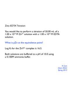
6 By the turn of the twenty-first century, the field of Fluorescence microscopy was responsible for a revolution in cell biology, coupling the power of live cell imaging to highly specific multiple labeling of individual organelles and macromolecular complexes with synthetic and genetically encoded fluorescent probes. Timescale Range for Fluorescence Processes Transition Process Rate Constant Timescale (Seconds) S(0) => S(1) or S(n) Absorption ( Excitation )Instantaneous10-15 S(n) => S(1)Internal Conversionk(ic)10-14 to 10-10 S(1) => S(1) Vibrational Relaxationk(vr)10-12 to 10-10 S(1) => S(0) Fluorescencek(f) or 10-9 to 10-7 S(1) => T(1) Intersystem Crossingk(pT)10-10 to 10-8 S(1) => S(0) Non-Radiative RelaxationQuenching k(nr), k(q) 10-7 to 10-5 T(1) => S(0) Phosphorescencek(p)10-3 to 100 T(1) => S(0) Non-Radiative RelaxationQuenching k(nr), k(qT)
7 10-3 to 100 Table 1 Fluorescence is generally studied with highly conjugated polycyclic aromatic molecules that exist at any one of several energy levels in the ground state, each associated with a specific arrangement of electronic molecular orbitals. The electronic state of a molecule determines the distribution of negative charge and the overall molecular geometry. For any particular molecule, several different electronic states exist (illustrated as S(0), S(1), and S(2) in Figure 1), depending on the total electron energy and the symmetry of various electron spin states.
8 Each electronic state is further subdivided into a number of vibrational and rotational energy levels associated with the atomic nuclei and bonding orbitals. The ground state for most organic molecules is an electronic singlet in which all electrons are spin-paired (have opposite spins). At room temperature, very few molecules have enough internal energy to exist in any state other than the lowest vibrational level of the ground state, and thus, Excitation processes usually originate from this energy level. The category of molecules capable of undergoing electronic transitions that ultimately result in Fluorescence are known as fluorescent probes, fluorochromes, or simply dyes.
9 Fluorochromes that are conjugated to a larger macromolecule (such as a nucleic acid, lipid, enzyme, or protein) through adsorption or covalent bonds are termed fluorophores. In general, fluorophores are divided into two broad classes, termed intrinsic and extrinsic. Intrinsic fluorophores, such as aromatic amino acids, neurotransmitters, porphyrins, and green fluorescent protein, are those that occur naturally. Extrinsic fluorophores are synthetic dyes or modified biochemicals that are added to a specimen to produce Fluorescence with specific spectral properties. Absorption, Excitation , and Emission Absorption of energy by fluorochromes occurs between the closely spaced vibrational and rotational energy levels of the excited states in different molecular orbitals.
10 The various energy levels involved in the absorption and Emission of light by a fluorophore are classically presented by a Jablonski energy diagram (see Figure 1), named in honor of the Polish physicist Professor Alexander Jablonski. A typical Jablonski diagram illustrates the singlet ground (S(0)) state, as well as the first (S(1)) and second (S(2)) excited singlet states as a stack of horizontal lines. In Figure 1, the thicker lines represent electronic energy levels, while the thinner lines denote the various vibrational energy states (rotational energy states are ignored).