
EdU (5-ethynyl-2’-deoxyuridine)
Fluorescence microscopy (Imaging) 50 coverslips Alexa Fluor® 488 dye C10337 • Includes blue-fluorescent nuclear counterstain, Hoechst 33342 • Not interchangeable with flow cytometry assays Alexa Fluor® 555 dye C10338 Alexa Fluor® 594 dye C10339 Alexa Fluor® 647 dye C10340 Table 4. Dye- and hapten-containing azides.
Download EdU (5-ethynyl-2’-deoxyuridine)
Information
Domain:
Source:
Link to this page:
Documents from same domain
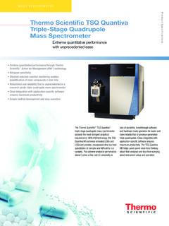
Thermo Scienti˜ c TSQ Quantiva Triple-Stage Quadrupole ...
tools.thermofisher.comThermo Scienti˜ c TSQ Quantiva Triple-Stage Quadrupole Mass Spectrometer ... • Thermo Scientifi c ... New Zealand +64 9 980 6700 Norway +46 8 556 468 00 Russia/CIS +43 1 333 50 34 0 Singapore +65 6289 1190 Spain +34 914 845 965 Sweden +46 8 556 468 00 Switzerland +41 61 716 77 00
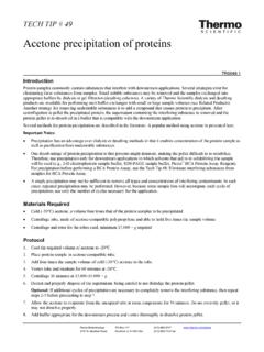
Acetone precipitation of proteins - Thermo Fisher Scientific
tools.thermofisher.com• Precipitation has an advantage over dialysis or desalting methods in that it enables concentration of the protein sample as well as purification from undesirable substances. • One disadvantage of protein precipitation is that proteins might denature, making the pellet difficult to re-solubilize.
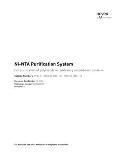
Ni-NTA Purification System - Thermo Fisher Scientific
tools.thermofisher.com7 Introduction Overview Introduction The Ni-NTA Purification System is designed for purification of 6xHis-tagged recombinant proteins expressed in bacteria, insect, and mammalian cells. The system is designed around the high affinity and selectivity of Ni-NTA Agarose
TRIzol Reagent User Guide - Pub. no. MAN0001271 - Rev. A
tools.thermofisher.comTRIzol™ Reagent Catalog Numbers 15596026 and 15596018 Doc. Part No. 15596026.PPS Pub. No. MAN0001271 Rev. A.0 WARNING! Read the Safety Data Sheets (SDSs) and follow the
Interpaion IEnPp fR nmpmeft n CfhRn Labware Chemical ...
tools.thermofisher.com286 www.thermoscientifi c.com Labwre Olafira nOPao ymreblo LsFoOrao LuapeDisEHLFbDabwriweFb CMT TTLy TL TM TT T/C T/ / ewFb TT na as H MnCM TM TTM Chemhical,l contaroAcSnlno#E N,o u u b u b b M M M b u u .
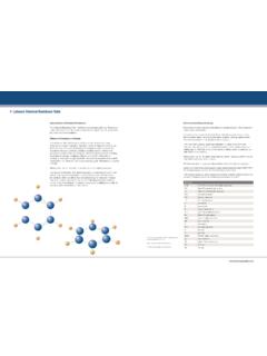
Interpretation of Nucleic Acid 260/280 Ratios
tools.thermofisher.comT123 – TECHNICAL BULLETIN NanoDrop Lite Interpretation of Nucleic Acid 260/280 Ratios T123– Rev 1/2012 Thermo Scientific NanoDrop Products Wilmington, Delaware USA Technical support:[email protected]
Corona CAD Charged Aerosol Detector - Thermo Fisher …
tools.thermofisher.com3 Corona CaD DeteCtor SpeCifiCationS* Operating Mode Charged Aerosol Detection Mobile Phase Flow Rate Up to 2 mL/min Full Scale Output Range 1 pA to 470 pA
GeneAmp PCR System 9700 - Thermo Fisher Scientific
tools.thermofisher.comIntroduction and Safety 1-1 Introduction and Safety 1 Overview About This Chapter This chapter provides information to help you safely operate the GeneAmp PCR System 9700. In This Chapter The following topics are covered in this chapter: Topics See Page About This Manual 1-2
alamarBlue TI read only - Thermo Fisher Scientific
tools.thermofisher.comPage 1 of 27 alamarBlue® Assay U.S. Patent No. 5,501,959 Indications for Use The alamarBlue® Assay is designed to measure quantitatively the proliferation of various human and animal cell lines, bacteria and fungi. The bioassay may also be used to establish relative

Installation Guide -Chromeleon 7 - Thermo Fisher Scientific
tools.thermofisher.comNote: Indicates information of special interest. Tip: Indicates information that will help you to use the software more efficiently. 1.3 Other Documents
Related documents
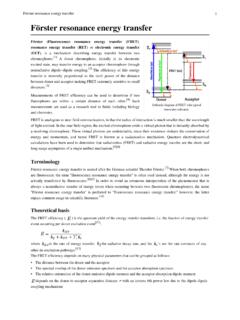
Förster resonance energy transfer
atlas.physics.arizona.eduIn fluorescence microscopy, fluorescence confocal laser scanning microscopy, as well as in molecular biology, FRET is a useful tool to quantify molecular dynamics in biophysics and biochemistry, such as protein-protein interactions, protein–DNA interactions, and protein conformational changes. For monitoring the complex formation
Fundamentals of Scanning Electron Microscopy and Energy ...
nanotechftm.tmf.bg.ac.rsNFMC Spring School on Electron Microscopy, April 2011 FWHM2=kE+FWHM noise 2 XEDS: Artifacts Si “escape peak” † 1.74keV bellow true characteristic peak position NFMC Spring School on Electron Microscopy, April 2011 The internal fluorescence peak † A small Si KDpeak at 1.74keV The sum peak † too high count rate

Characteristics of Benign & Malignant Neoplasms
www.columbia.edu-light microscopy: biopsy-cytology (Fine Needle Aspiration—FNA)-immunohistochemistry-fluorescence in situ hybridization (FISH)-molecular probes, incl. gene microarray-flow cytometry (lymphomas, leukemias) 10 Tumor Markers *Molecules in plasma produced by tumor cells

Polymer-Modified Asphalt 101
pavement.engineering.asu.eduNov 12, 2014 · Fluorescence Microscopy Optical reflective fluorescence photomicrograph of a swollen polymer-asphalt blend. Initial Dispersion of Polymer in Asphalt . Fluorescence Microscopy Formation of Polymer Networks in Asphalt . Fluorescence Microscopy Maturing of Polymer Network in Asphalt .
MitoTracker® Mitochondrion-Selective Probes
tools.thermofisher.comanalysis, or fluorescence microscopy. If immobilized cells on coverslips are needed, use poly-D-lysine to coat the slides or coverslips before mounting. If the cells are to be fixed and permeabil ized, continue to Fixation and Permeabilization after Staining. Optional: Fixation and Permea-

Fluorescence Excitation and Emission Fundamentals
www.chem.uci.eduFluorescence was first encountered in optical microscopy during the early part of the twentieth century by several notable scientists, including August Köhler and Carl Reichert, who initially reported that fluorescence was a nuisance in ultraviolet microscopy.
GRAPHENE OXIDE DETECTION IN AQUEOUS SUSPENSION
www.thecompleteguidetohealth.coma) Bright field. b-d) Fluorescence extinction microscopy (FQM) Kim et al, 2010. Seeing graphene-based sheets, Materials Today, Volume 13, 2010, Pages 28-38, Literature Image Low magnification TEM "The figure shows a TEM image of bilayer graphene with edges that tend to curl and bend slightly" 5

The Handbook - Laboratory Diagnosis of Tuberculosis by ...
www.who.intmicroscopy of two consecutive sputum specimens will identify the vast majority (95–98%) of smear-positive TB patients**. Moreover, microscopy can be decentralised to peripheral laboratories. Despite its advantages sputum smear microscopy does fall short in test sensitivity, especially for certain patient groups such as those living