Transcription of An introduction to the anatomy of the uterine cervix
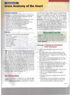
1 1 Chapter 1An introduction to the anatomy of the uterine cervixA thorough understanding of the anatomy andphysiology of the cervix is absolutely essential foreffective colposcopic practice. This chapter deals withthe gross and microscopic anatomy of the uterine cervixand the physiology of the transformation zone. Thecervix is the lower fibromuscular portion of the is cylindrical or conical in shape, and measures 3 to 4cm in length, and cm in diameter. It is supported bythe cardinal and uterosacral ligaments, which stretchbetween the lateral and posterior portions of the cervixand the walls of the bony pelvis. The lower half of thecervix, called the portio vaginalis, protrudes into thevagina through its anterior wall, and the upper halfremains above the vagina (Figure ).
2 The portiovaginalis opens into the vagina through an orifice calledthe external os. The cervix varies in size and shape depending onthe woman s age, parity and hormonal status. Inparous women, it is bulky and the external os appearsas a wide, gaping, transverse slit. In nulliparouswomen, the external os resembles a small circularopening in the centre of the cervix . The supravaginalportion meets with the muscular body of the uterusat the internal cervical os. The portion of the cervixlying exterior to the external os is called theectocervix. This is the portion of the cervix that is The cervix , the lower fibromuscular portion of the uterus, measures 3-4 cm in length and cmin diameter; however, it varies in size and shape depending on age, parity and menstrual status ofthe woman.
3 Ectocervix is the most readily visible portion of the cervix ; endocervix is largely invisible and liesproximal to the external os. Ectocervix is covered by a pink stratified squamous epithelium, consisting of multiple layers of cellsand a reddish columnar epithelium consisting of a single layer of cells lines the endocervix. Theintermediate and superficial cell layers of the squamous epithelium contain glycogen. The location of squamocolumnar junction in relation to the external os varies depending upon age,menstrual status, and other factors such as pregnancy and oral contraceptive use. Ectropion refers to the eversion of the columnar epithelium onto the ectocervix, when the cervixgrows rapidly and enlarges under the influence of estrogen, after menarche and during pregnancy.
4 Squamous metaplasia in the cervix refers to the physiological replacement of the everted columnarepithelium on the ectocervix by a newly formed squamous epithelium from the subcolumnarreserve cells. The region of the cervix where squamous metaplasia occurs is referred to as the transformation zone. Identifying the transformation zone is of great importance in colposcopy, as almost all manifestationsof cervical carcinogenesis occur in this vaginalisEndocervical canalCervixBladderAnterior fornixPubic boneUrethrareadily visible on speculum examination. The portionproximal to the external os is called the endocervixand the external os needs to be stretched or dilatedto view this portion of the cervix . The endocervicalcanal, which traverses the endocervix, connects theuterine cavity with the vagina and extends from theinternal to the external os, where it opens into thevagina.
5 It varies in length and width depending on thewoman s age and hormonal status. It is widest inwomen in the reproductive age group, when itmeasures 6-8 mm in space surrounding the cervix in the vaginal cavityis called the vaginal fornix. The part of the fornixbetween the cervix and the lateral vaginal walls is called2 Chapter 1 FIGURE : gross anatomy of the uterine cervixFundusFallopian tubeBody of uterusSupravaginal cervixInternal osEndocervixLateral fornixExternal osEctocervixVaginaUterusPosterior fornixRectumSacrumVagina3the lateral fornix; the portions between the anterior andposterior walls of the vagina and the cervix are termedthe anterior and posterior fornix, stroma of the cervix is composed of dense,fibro-muscular tissue through which vascular,lymphatic and nerve supplies to the cervix pass andform a complex plexus.
6 The arterial supply of thecervix is derived from internal iliac arteries throughthe cervical and vaginal branches of the uterinearteries. The cervical branches of the uterine arteriesdescend in the lateral aspects of the cervix at 3 and9 o clock positions. The veins of the cervix runparallel to the arteries and drain into the hypogastricvenous plexus. The lymphatic vessels from the cervixdrain into the common, external and internal iliacnodes, obturator and the parametrial nodes. Thenerve supply to the cervix is derived from thehypogastric plexus. The endocervix has extensivesensory nerve endings, while there are very few inthe ectocervix. Hence, procedures such as biopsy,electrocoagulation and cryotherapy are welltolerated in most women without local sympathetic and parasympathetic fibres arealso abundant in the endocervix, dilatation andcurettage of the endocervix may occasionally lead toa vasovagal reaction.
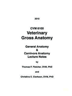
7 The cervix is covered by both stratified non-keratinizing squamous and columnar two types of epithelium meet at thesquamocolumnar junction. Stratified non-keratinizing squamousepitheliumNormally, a large area of ectocervix is covered by astratified, non-keratinizing, glycogen-containingsquamous epithelium. It is opaque, has multiple (15-20)layers of cells (Figure ) and is pale pink in colour. Thisepithelium may be native to the site formed duringembryonic life, which is called the native or originalsquamous epithelium, or it may have been newlyformed as metaplastic squamous epithelium in earlyadult life. In premenopausal women, the originalsquamous epithelium is pinkish in colour, whereas thenewly formed metaplastic squamous epithelium lookssomewhat pinkish-white on visual examination.
8 The histological architecture of the squamousepithelium of the cervix reveals, at the bottom, asingle layer of round basal cells with a large dark-staining nuclei and little cytoplasm, attached to thebasement membrane (Figure ). The basementmembrane separates the epithelium from theunderlying stroma. The epithelial-stromal junction isusually straight. Sometimes it is slightly undulatingwith short projections of stroma at regular stromal projections are called papillae. Theparts of the epithelium between the papillae are calledrete pegs. The basal cells divide and mature to form the nextfew layers of cells called parabasal cells, which alsohave relatively large dark-staining nuclei andgreenish-blue basophilic cytoplasm.
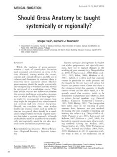
9 FurtherAn introduction to the anatomy of the uterine cervixFIGURE :Stratified squamous epithelium (x 20)Superficial cell layerParabasal layerBasal cell layerIntermediate cell layerStromalpapillaStromaBasement membrane4 Chapter 1differentiation and maturation of these cells leads tothe intermediate layers of polygonal cells withabundant cytoplasm and small round nuclei. Thesecells form a basket-weave pattern. With furthermaturation, the large and markedly flattened cellswith small, dense, pyknotic nuclei and transparentcytoplasm of the superficial layers are , from the basal to the superficial layer, thesecells undergo an increase in size and a reduction ofnuclear size. The intermediate and superficial layer cellscontain abundant glycogen in their cytoplasm, whichstains mahogany brown or black after application ofLugol s iodine and magenta with periodic acid-Schiffstain in histological sections.
10 Glycogenation of theintermediate and superficial layers is a sign of normalmaturation and development of the squamousepithelium. Abnormal or altered maturation ischaracterized by a lack of glycogen production. The maturation of the squamous epithelium of thecervix is dependent on estrogen, the female estrogen is lacking, full maturation andglycogenation does not take place. Hence, aftermenopause, the cells do not mature beyond theparabasal layer and do not accumulate as multiplelayers of flat cells. Consequently, the epitheliumbecomes thin and atrophic. On visual examination, itappears pale, with subepithelial petechialhaemorrhagic spots, as it is easily prone to trauma.