Transcription of Gross Anatomy of the Brain
1 Note that the clinical case at the beginning of the chapterrefers to a patient who loses consciousness subsequent tohead is it important to do a CT scan or MRI in the case of a head injurythat involves loss of consciousness?2 What happened to the patient s Brain as his head was struck by thesteel beam?3 Most tissues swell when they are exposed to blunt trauma. Does thathappen to the Brain ?A 28-year-old man was struckin the back of the head by a steelbeam while at work. He apparently lostconsciousness for approximately 30 minutes. He was hospitalized since hewas still confused and disorientedthroughout his stay in the emergencyroom. By the next day he was felt to beback to normal except for a headache and was discharged. The patient had nomemory of the event, and he had nomemory of an important conversation hehad had with his boss 5 minutes beforethe head trauma. He also had no memoryof being in the emergency room and nomemories of events subsequent to theinjury until the next day.
2 He also noted,besides persistent headache, that he was somewhat forgetful since the injury. Neurologic examination was CASEThe Brain , a bilaterally symmetric, soft, gelatinous struc-ture surrounded by its meninges and enclosed in its bony cranium, is continuous with the spinal cord at the foramenmagnum at the base of the skull. At birth the Brain weighsless than 400 g, but by the beginning of the second year of lifeit has more than doubled in weight to 900 g. The adult brainweighs between 1,250 and 1,450 g, and demonstrates a gen-der differential, since brains of males generally weigh morethan those of females. This statement, however, should betempered by the evidence that, in adults, the ratio of Brain tobody weight is greater in females than in males and that theincrease in weight is due more to the proliferation of neuro-glia than to the mitotic activity of neurons. An additionalpoint of interest is that there does not appear to be a relation-ship between Brain weight and detailed in Chapter 2, it is evident during embryo-genesis that the Brain is subdivided into five continuousregions, from rostral to caudal: the telencephalon, dienceph-alon, mesencephalon, metencephalon, and the Brain grows in size and complexity, these regions fold upon and over one another, so that in the adult the evidence of these subdivisions is no longer clearly present chapter will not discuss the functional aspectsof the Brain .
3 Instead its basic morphology and architectureCHAPTER 6 Gross Anatomy of the BrainCLINICAL CASECEREBRUMDIENCEPHALONCEREBELLUMBRAINS TEMCLINICAL CONSIDERATIONSSYNONYMS AND EPONYMSFOLLOW-UP TO CLINICAL CASEQUESTIONS TO PONDERATOC06 3/17/06 9:59 AM Page 68are detailed to provide an anatomical framework of referencefor the chapters that follow, and many of the major topicsintroduced in this chapter are discussed in further detail inspecific chapters in this textbook. Much of this terminologyshould be memorized so that when, in later chapters, func-tional connections among regions of the Brain are discussedthe student has a visual image of the location of the variousstructures and the pathways the connections the adult Brain is viewed in three dimensions, only threeregions are clearly visible, and these are the cerebrum, cere-bellum, and part of the cerebral hemispheresare narrower posteriorly, atthe occipital pole, than an-teriorly, at the frontal are large, oval struc-tures that superficially resemble the surface of a shelled walnut (Fig.)
4 The midline longitudinal cerebral fissure,occupied in life by the falx cerebri, incompletely separates thetwo cerebral hemispheres from one another. The floor of thecerebral fissure is formed by the corpus callosum, a largemyelinated fiber tract that forms an anatomical and func-tional connection between the right and left surface few millimeters of the cerebral hemisphere arecomposed of a highly folded collection of gray matter, knownas the cerebral cortex. This folding increases the surface areaand presents elevations, gyri, and depressions, sulci. Deep tothe cortex is a central core of white matterthat forms the bulkof the cerebrum and represents fiber tracts, supported byneuroglia, ferrying information destined for the cortex andcortical responses to other regions of the central nervous system (CNS). Buried within the mass of white matter are collections of neuron cell bodies, some of which are lumpedtogether under the rubric of basal ganglia, even though, tech-nically, they are nuclei.
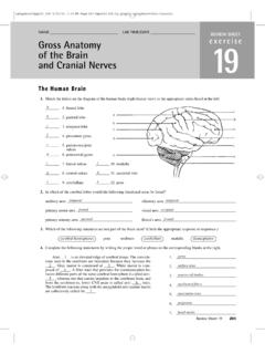
5 Large collections of gray matter areGROSS Anatomy OF THE Brain ===69 The cerebrum, observed from above,hides the remainder of the Brain fromview, and is composed of two large,oval, cerebral hemispheresCentral sulcusPrecentral gyrus (motor area)Postcentral gyrus (sensory area)Sensory speech areaAngular gyrusSuperior parietal lobuleOccipital lobeCerebellumPonsMedulla oblongataParietal lobeLateral sulcus (fissure)Superior temporal gyrusMiddle temporal gyrusInferior temporal gyrusSupramarginal gyrusIntraparietal sulcusParieto-occiptal sulcusPostcentral sulcusPrecentral sulcusFrontal lobeMotor speech areaTemporal lobeInferior frontalgyrusMiddle frontal gyrusSuperior frontal gyrusPreoccipital notchFigure of the Brain from a lateral 3/17/06 9:59 AM Page 69also present in the diencephalons, namely, the epithalamus,thalamus, hypothalamus, and cerebrumis a hollow structure and the cavities withinthe cerebral hemispheres are called the right and left lateralventricles, which communicate with the third ventricle viathe interventricular foramen(foramen of Monro) (Fig.)
6 The two lateral ventricles are separated from one another bytwo closely adjoined non-nervous membranes, each knownas a septum pellucidum. Ependymal cells line each lateralventricle, and protruding into each ventricle is a choroid plexusthat functions in the manufacture of cerebrospinal of the cerebral hemispheresEach cerebral hemisphere issubdivided into five lobes:the frontal, parietal, tem-poral, and occipital lobes,and the insula (Table ).Additionally, the cortical constituents of the limbic systemare also considered to be a region of the cerebral hemisphereand some consider it to be the sixth lobe, the limbic from the side, each cerebral hemisphere re-sembles the shape of a boxing glove, where the thumb is the temporal lobeand is separated from the parietal lobeby the lateral fissure(fissure of Sylvius) (Fig. ). The floor ofthe lateral fissure is formed by the insula(island of Reil) thatis hidden by the frontal, parietal, and temporal opercula(L.)
7 , lids ), regions of the same named lobes. Although the geographic distributions of many of the sulci and gyri are relatively inconsistent, some regularly occupy specific locations, are recognizable in most brains, and are sulci are generally smaller and shallower than thefissures, and one of these, the central sulcus(central sulcus ofRolando), separates the frontal lobe from the parietal division between the parietal and occipital lobes is notreadily evident when viewed from the lateral aspect becauseit is defined as the imaginary line between the preoccipitalnotchand the parieto-occipital notch. However, it is clearlydelimited on the medial aspect of the cerebral hemisphere,where the boundary between these two structures is theparieto-occipital sulcusand its continuation, the calcarinefissure (Fig. ).Frontal lobeThe frontal lobe extends from the frontal pole to the central sulcusOn its lateral aspect, the frontal lobeextends from the frontalpole to the central sulcus, constituting the anterior one-thirdof the cerebral cortex.
8 Its posteriormost gyrus, theprecentralgyrus, consists of the primary motor area and is borderedanteriorly by the precentral sulcusand posteriorly by the70===CHAPTER 6 The five lobes of the cerebralhemispheres are the frontal, parietal,temporal, and occipital lobes, and theinsulaLobeFrontalParietalTemporalOcci pitalInsulaLimbicTable of the cerebral gyriPrecentral gyrusInferior frontal gyrusAnterior paracentral lobuleGyrus rectus and orbital gyriPostcentral gyrusSuperior parietal lobuleInferior parietal lobuleSupramarginal gyrusAngular gyrusPosterior paracentral lobule precuneusSuperior temporal gyrusMiddle and inferior temporal gyriTransverse temporal gyri (of Heschl)Fusiform gyrusSuperior and inferior occipital gyriCuneate gyrus (cuneus)Lingual gyrusShort and long gyriCingulate gyrusParahippocampal gyrusHippocampal formationSubcallosal, parolfactory, andpreterminal gyriFunction/commentPrimary motor areaBroca s area in dominant hemisphere; functions in speech productionContinuation of precentral gyrusOlfactory bulb and tract in the olfactory sulcusPrimary somesthetic areaAssociation area involved in somatosensory functionIntegrates auditory, visual, and somatosensory informationReceives visual inputContinuation of the postcentral gyrusWernicke s area in dominant hemisphere; ability to read, understand, and speak the written wordPrimary auditory cortexBorders the parahippocampal gyrus of the limbic lobeSeparated from each other by the lateral occipital sulcusSeparated from each other by the calcarine fissure; striate cortex (primary visual cortex) is on the banks of this fissureForms the floor of the lateral sulcus; associated with tasteAbove the body of the corpus callosum and continues as the isthmusAnterior continuation of isthmus.
9 Ends in the uncusComposed of hippocampus, subiculum, and dentate gyrusCollectively known as the subcallosal areaATOC06 3/17/06 9:59 AM Page 70 Gross Anatomy OF THE Brain ===71 Anterior hornInferior hornBodyTrigoneThird ventricleCerebralaqueductPosterior hornFourth ventricleLateral apertureLateral ventricleLeft and right ventriclesInterventricular foramenThird ventricleCerebral aqueductFourth ventricleMedian aperture of MagendieLateral aperturesof LuschkaBACentral canalSubarachnoid spaceArachnoid granulationsSuperior sagittal sinusGreat cerebral veinStraight sinusConfluence of sinusesFigure (A) Diagram of the ventricles of the Brain and central canal of the spinal cord in situ. (B) A three-dimensional representation of the ventricles of the 3/17/06 9:59 AM Page 71central sulcus. The region of the frontal lobe located anteriorto the precentral sulcus is subdivided into the superior, middle, and inferior frontal gyri. This subdivision is due to the presence, though inconsistent, of two longitudinallydisposed sulci, the superior and inferior frontal sulci.
10 Theinferior frontal gyrus is demarcated by extensions of the lat-eral fissure into three subregions: the pars triangularis, parsopercularis, and pars orbitalis. In the dominant hemisphere, aregion of the inferior frontal gyrus is known as Broca s area,which functions in the production of its inferior aspect, the frontal lobe presents the longitu-dinally disposed olfactory sulcus. Medial to this sulcus is thegyrus rectus (also known as the straight gyrus), and lateral toit are the orbital gyri. The olfactory sulcus is partly occupiedby the olfactory bulband olfactory tract (Figs , ). At its posterior extent, the olfactory tract bifurcates to form the lateral and medial olfactory striae. The intervening area be-tween the two striae is triangular in shape and is known asthe olfactory trigoneand it abuts the anterior its medial aspect, the frontal lobe is bordered by thearched cingulate sulcus, which forms the boundary of the sup-erior aspect of the cingulate gyrus.