Transcription of Exercise 20 Gross Anatomy of the Heart - Oxford …
1 Exercise 20 Gross Anatomy of the Heart Laboratory Objectives On completion of the activities in this Exercise , you "'rill be able to: Describe the anatomical relations of the Heart with other structures in the thoracic cavity. Provide details on the arrangement of the connective tissue layers (the pericardium) that surround the Heart . Identify important structures on the surface of the Heart . Locate the major internal structures of the Heart . Identify the tissue layers of the Heart wall. Describe the coronary circulation. Describe the flow of blood through the Heart . Dissect a sheep Heart and compare its structure with the human Heart .
2 Materials Anatomical models or figures of the human Heart Plastic bags Cotton or cheesecloth Colored pencils Preserved sheep hearts Dissecting trays Dissecting tools Dissecting gloves Protective eyewear Face mask The Heart is a two-sided, double-pumping organ. The left side (the left pump) controls the flow of blood to all tissues and cells in the body, where oxygen and nutrients are delivered and metabolic wastes are taken away. The right side (the right pump) sends blood to the lungs, where oxygen stores in red blood cells are replenished and carbon dioxide, a metabolic waste, is re leased. To keep blood circulating throughout the body, the Heart beats approximately 100,000 times and pumps between 7000 and 9000 liters of blood each day.
3 By any standard, this is an arduous workload, but the fact that the Heart can maintain this level of ac ti\-ity for decades, without stopping, is nothing short of remarkable. In this Exercise , you will examine the special anatomical fea tures that reflect the enduring and efficient functioning of the Heart . You will focus your examination on Gross anatomical lructure. If you would like to review the light microscopic st ru 'LUre of cardiac muscle, see Activity The Pericardium The Heart is enclosed by a membranous sac called the peri cardium (Figure ) . This structure consists of two parts. The outer fibrous pericardium is a tough, fibrous connective tissue layer that is fused to adjacent structures (the diaphragm, sternum, costal cartilages of ribs, thoracic vertebrae, and the great vessels emerging from the Heart ).
4 The inner serous peri cardium is a delicate serous membrane that forms a double layered sac around the Heart . It consists of the parietal pericardium, which covers the deep or inner surface of the fi brous pericardium, and the visceral pericardium, which forms the outer surface of the Heart wall. The potential space between the parietal and visceral pericardial membranes is the pericardial cavity (Figure ). The cavity is filled with a wa tery fluid produced by the epithelial cells lining the serous peri cardium. The fluid helps to reduce friction when the two serous membranes rub against each other as the Heart beats. CLINICAL CORRELATION Inflammation of the pericardiaI membranes, known as peri carditis, increases the friction between the two membranes and causes an overproduction of fluid.
5 As the fluid accumulates in the pericardial cavity it inhibits the normal movements of the Heart wall and restricts cardiac output, leading to a condition called cardiac tamponade. ACTIVITY Examining the Organization of the Pericardium 1. Obtain a large, clear plastic bag and close off the open end. 2. With a Heart model, push the inferior pOinted tip (the apex of the Heart ) into the wall of the closed plastic bag. This action is similar to pushing a fist into the bag as illus trated in Figure 3. Notice that as you push the Heart deeper into the closed bag, two layers of plastiC, separated by a space, cover the organ (Figure ).
6 4. Continue pushing the Heart into the bag untill you reach the great vessels that are attached to the superior aspect of the Heart (aorta, pulmonary trunk, superior vena cava). 5. The plastiC layers represent the serous pericardium (Figure ), as follows: The inner plastiC layer that is in contact with Heart wall represents the visceral pericardium. The outer layer of plastiC represents the parietal pericardium. The space between the two plastic layers is the pericar dial cavity. Notice that the two layers of plastiC are continuous with each other at the great vessels. In other words, the visceral pericardium is continuous with the 355 --------------------356 Exercise TWENTY parietal pericardium where the great vessels are con What important function does the fibrous peri nected to the Heart (Figure ).
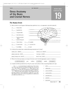
7 Cardium serve? 6. Wrap a layer of cotton or cheesecloth over the outer plas tic layer (the parietal pericardi u m). This layer represents the fibrous pericardium which is attached to the connec tive tissue layers that surround adjacent structures. Thyroid gland Right lung _ _ --First rib (cut) Aorta (segment removed) (a) Right pleural cavity Rig ht pulmonary -1:-11k-------.~f":::9:">C- ..__~~..;f artery Right pulmonary Base of Heart Pericardial cavity containing pericardial attachment to diaphragm fluid Cut edge of ~~ , , Fibrous ---'- vein Superior vena cava Right atrium Parietal pericardium parietal pericardium Fibrous tissue of w~pericardial sac Areolar tissue Mesothelium } Cut edge of epicardium (visceral pericardium) Apex of Heart (b) Parietal pericardium Outer wall (corresponds to parietal pericardium) Epicardium (visceral pericardium) Wrist (corresponds to base of Heart ) (c) Figure The anatomical relations of the Heart .
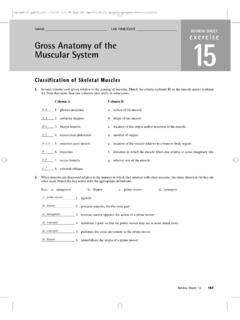
8 A) Anterior view of the thoracic cavity, showing the Heart within the mediastinum and between the two lungs; b) transverse section of the thoracic cavity showing the position of the Heart in relation to other structures; c) the relationship of the Heart , pericardium, and pericar dial cavity. Left pleural cavity Inner wall (corresponds to visceral pericardium) Air space (corresponds to pericardia I cavity) Balloon Superior Auricle of RIGHT ATRIUM Coronary sulcus RIGHT Pericardiu Gross A NATOMY OF THE Heart Arch of aorta Ligamentum arteriosum Descending aortaAscending --#!!!jii!~I! aorta Left pulmonary artery ___:--_ Pulmonary cava trunk Auricle of ---"jl--- right atrium Fat in anteriorFat in.
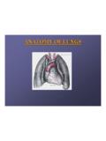
9 '--~,. interventricularcoronary sulcus \1I:1~ITiiHt'LE (a) Pericardium Ascending Pulmonary Auricle of trunk left atrium right atrium VENTRICLE to diaphragm Left pulmonary artery Left pulmonary ""':::::----:;;d'l!!!llllllll~ Fat in coronary sulcus Coronary sinus interventricular LEFT sulcus VENTRICLE (b) ~ ~.""'~Fat in posterior interventricular sulcus (e) e External Anatomy of the Heart . a) Diagram, and b) dissec --",~ :erior view; c) diagram, posterior view. Gross Anatomy of the Heart The h eart is a four-chambered organ that is shaped roughly like an inverted pear. On average, it is approximately 14 em long and 9 em wide, or slightly larger than a cl enched fist.
10 Its weight ranges from 230 to 280 grams in females and 280 to 340 grams in males. The h ea rt and its surrolU)ding pericardial cavi ty are lo cated within the mediastinum, a centrally located area within the thoracic cavity. Two thirds of the organ is pOSitioned to the left of the midline. It is bordered laterally by the pleural cavities, which surround the lungs, anteriorly by the sternum, and pos teriorly by the esophagus and thoracic vertebrae (F igures and b) ACTIVITY Examining the Gross Anatomy of tbe Human Heart External Anatomy 1. Examine a model of the Heart from an anterior view (Figures and b) and make the following observations.