Transcription of Cardiac Biomarkers - Open
1 2 Cardiac Biomarkers Sadip Pant1, Abhishek Deshmukh1, Pritam Neupane2, Kavin Kumar3 and Vijayashankar4 1 University of Arkansas For Medical Sciences, Little Rock, AR 2 Medical College of Georgia, Augusta, GA 3 Department of Internal Medicine, Priya Hospital-Heart and Diabetic Care Tamilnadu 4 Apollo Hospitals Greams Road, Chennai, Tamilnadu 1,2 USA 3,4 India 1. Introduction 1950 s: Clinical reports that transaminases released from dying myocytes could be detected via laboratory testing, aiding in the diagnosis of myocardial infarction. The race to define clinical markers to aid in the diagnosis, prognosis, and risk stratification of patients with potential cardiovascular disease begins. Initial serum markers included AST, LDH, total CK and -hydroxybutyrate. These enzymes are all released in varying amounts by dying myocytes.
2 Lack of sensitivity and specificity for Cardiac muscle necrosis fuels continued research. 1960 s:CK known to be released during muscle necrosis (including Cardiac ).Quantitative assays were cumbersome and difficult to perform. Total CK designed as a fast, reproducible spectrophotometric assay in the late 1960 isoenzymes are subsequently described: MM, MB and BB fractions. In 1970 s MB fraction noted to be elevated in and highly specific for acute MI. CKMB now measured via a highly sensitive monoclonal antibody assay. It was felt for a time that quantitative CKMB determination could be used to enzymatically measure the size of an infarct. This has been complicated by release of additional enzymes during reperfusion. As CK-MB assays become more sensitive, researchers come to the paradoxical realization that it too is not totally Cardiac specific.
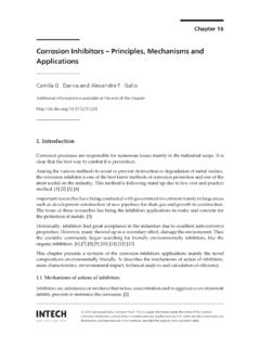
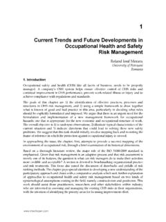
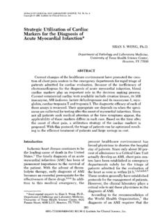
3 The MB fraction is determined to be expressed in skeletal muscle, particularly during the process of muscle regeneration and the search for Cardiac specificity continues. Research turns towards isolation of and development of assays for sarcomeric proteins. Myosin light chains were originally isolated and then subsequently abandoned because of specificity issues. Troponin I first described as a biomarker specific for AMI in 1987; Troponin T in 1989. Now troponins are the biochemical gold standard for the diagnosis of acute myocardial infarction via consensus of ESC/ACC. Novel Strategies in Ischemic Heart Disease 18 Fig. 1. Timeline showing landmark events in the development of Cardiac Biomarkers Cardiac Markers: What are we looking at? A biomarker is defined as a measurable substance or parameter that is an indicator of an underlying biological or pathological process.
4 Therefore, depending on the underlying process that we are referring to, the Cardiac markers can be classified as markers of necrosis, markers of ischemia and markers of inflammation. The features of an ideal Cardiac marker would be: High sensitivity and specificity Rise and fall rapidly after ischemia Able to perform reliably and uniformly Be simple to perform Have turnaround time <60 min Not influenced by functioning of other organs, in particular, functioning of kidney. Therefore, Cardiac marker is an umbrella term which is used to define present day used necrosis markers as well as all the upstream markers of necrosis studied/under study including proinflammatory cytokines, cellular adhesion molecules, acute phase reactants, plaque destabilization Biomarkers , plaque rupture biomarker and prenecrosis ischemia Biomarkers .
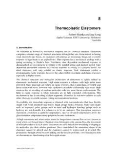
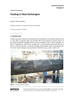
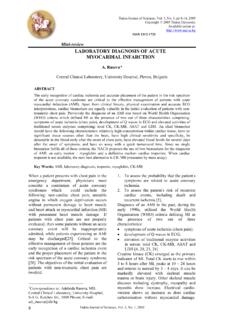
5 This can be simply visualized with the help of following flow diagram: Cardiac Biomarkers 19 Fig. 2. Flowchart showing significance of Biomarkers at various levels in the pathogenesis of acute coronary syndrome Markers of Cardiac necrosis Cardiac markers are used in the diagnosis and risk stratification of patients with chest pain and suspected acute coronary syndrome (ACS). The Cardiac troponins, in particular, have become the Cardiac markers of choice for patients with ACS. Indeed, Cardiac troponin is central to the definition of acute myocardial infarction (MI) in the consensus guidelines from the American College of Cardiology (ACC) and the European Society of Cardiology. Older Definition of Myocardial Infarction- WHO 1979 1. Definite acute myocardial infarction-Definite acute myocardial infarction is diagnosed in the presence of unequivocal EKG changes and/or unequivocal enzyme changes; the history may be typical or atypical.
6 2. Possible acute myocardial infarction-Possible acute myocardial infarction is diagnosed when serial, equivocal ECG changes persist more than 24 hours, with or without equivocal enzyme changes; the history may be typical or atypical. 3. Old myocardial infarction-Old myocardial infarction is usually diagnosed on an unequivocal ECG in the absence of a history or enzymatic signs of acute myocardial Proinflammatory Cytokines Plaque Destabilization Plaque Rupture Ischemia Necrosis hsCRP, Lp-PLA2, Hohomocysteine Matrix Metalloproteinases, Metal Independent Myeloperoxidases Soluble CD40 Lingand, placental growth factor, pregnancy associated plasma protein A Unbound Free Fatty Acids, Plasma Choline and Whole Blood Choline, Ischemia modified albuminCKMB, Cardiac Troponins, Myoglobin Novel Strategies in Ischemic Heart Disease 20infarction.
7 If there are no residual ECG changes, the diagnosis may be based on earlier, typical ECGs or on the presence of prior unequivocal serum enzyme changes. Redefinition of Myocardial Infarction- Joint Task Force of the European Society of Cardiology, American College of Cardiology Foundation, the American Heart Association, and the World Health Federation (ESC/ACCF/AHA/WHF) 2007 A typical rise and/or gradual fall (troponin) or more rapid rise and fall (CK-MB) of biochemical markers of myocardial necrosis, with at least one of the following is required: Ischemic symptoms Development of pathologic Q waves on the ECG ECG changes indicative of ischemia (ST segment elevation or depression) Imaging evidence of new loss of viable myocardium or a new regional wall motion abnormality. In addition, pathologic findings (generally at autopsy) of an acute MI are accepted criteria.
8 Markers of Cardiac necrosis have come a long way since 1950s. Some of the markers used in the past are no longer in use today. Current markers and those used in the past have been outlined in the table below. Those used in the past are not discussed separately further. Current Cardiac Markers CK-MB Myoglobin CKMB Isoforms Troponin I and T Cardiac Markers of the Past Total CK Activity Aspartate Aminotransferase Activity Lactate Dehydrogenase Activity LD1/LD2 Ratio Table 1. Various present day and past Cardiac Biomarkers 2. Creatine kinase The enzyme creatinine kinase (formerly referred to as creatinine phosphokinase) exists as three isoenzyme forms: CK-MM, CK- MB, and CK-BB. These isoenzymes are found in the cytosol and facilitate the egress of high energy phosphates into and out of mitochondria.
9 CK: Dimer composed of 2 monomers: M (43,000 Da) and B (44,500 Da)---- > CK BB or CK MB or CK MM Role: Creatine + ATP <---> ADP + Phosphocreatine + Energy (muscular contraction) CK BB : Increased in neurological diseases ; prostatectomy; digestive cancers CK MB : Increased with AMI CK MM :Increased in myopathy, hypothyroidism, polymyositis, rhabdomyolysis, muscle trauma, intensive exercise, AMI Table 2. Isoenzymes of Creatinine Kinase Distribution of CK: Creatine Kinase (CK) isoenzyme activity is distributed in a number of tissues. The percentage of CK-MB fraction found in the heart is higher than in most other tissues. However, sensitive radioimmunoassays are able to detect small amounts of B Cardiac Biomarkers 21 chain protein in skeletal muscle, and some muscles have been reported to contain up to 10 percent B chain protein. Most muscles have much more CK per gram than heart tissue.
10 As a result, despite containing only a small percent of B chain protein, skeletal muscle breakdown can lead to absolute increases in CK-MB in the plasma. Therefore, skeletal muscle damage can confound the diagnosis of an MI, as CK-MB can be released. The following are examples: Myocardial injury after cardiopulmonary resuscitation Cardioversion Defibrillation Cardiac and non- Cardiac surgical procedures Blunt chest trauma with possible Cardiac contusion Cocaine abuse Total CK, CK-MB and CK-MB to Total CK ratio: Since CK is widely distributed in tissues, elevations in total serum CK lack specificity for Cardiac damage, which improves with measurement of the MB fraction. The normal range of CK also varies considerably; a twofold or greater increase in the CK concentration is required for diagnosis.