Transcription of Ch. 14: Cardiovascular Physiology - people.fmarion.edu
1 1 Ch. 14: Cardiovascular Physiology 1. Objectives: 1. Review: Heart Anatomy, and Systemic & Pulmonary circuits. 2. The Cardiac Cycle and Heart Sounds 3. The Heart s Conduction Cycle & the ECG 4. Regulation of Heart s Pacemaker (heart rate) 5. Blood Pressure 6. Cardiac output and its Regulation 7. Three Ways the Body Regulates Blood Pressure 8. Abnormal Blood Pressure 9. Congestive Heart Failure 10. Blood Physiology 2. Anatomy of heart (Review go over QUICKLY) 3. Circulatory Circuits (Anatomy review go over QUICKLY) > Systemic circuit from L atrium, arteries, tissues, veins, to vena cava > Pulmonary circuit from R atrium, pulmonary arteries, lungs, and pulmonary veins. 4. Cardiac cycle =& sounds > Ventricular systole = when both L & R atria close (after they contracted to push blood into ventricles, the valves snap shut Lub or S1 sound.)
2 At this point the ventricles contract & push blood into pulmonary trunk & aorta > Ventricular diastole = ventricles relax, pulmonary & aortic semilunar valves snap shut, producing Dub or S2 sound. > Asystole = no heart sounds 5. Heart Defects (see online clinical app) > Heart Murmers: - Innocent murmur = quiet whoosh sound between S1 & S2 sounds. Can be normal in children (they outgrow it) BUT pathology if adult develops a murmur. - Pathological murmur = loud whoosh sound between S1 & S2. > could be valves not closing completely, leading to regurgitant flow (backflow of blood) > aortic stenosis (stiffening of aortic valve) could lead to it > rheumatic heart disease (autoimmune attack on mitral valve) can lead to it. > Septal Defects (these are before baby is born.)
3 TheseTholes supposed to close by birth. Not all do so.) 1) Patent foramen ovale (patent means open) = fetal hole between L & R atria 2) Patent ductus arteriosis = fetal hole between pulmonary trunk & aorta 3) Ventricular septal defect = fetal hole in interventricular septum 6. Review slide 7. Heart s Conduction System 1. SA (sinoatrial) node = heart s pacemaker in upper R atrium. Where an action potential spontaneous happens. Signal jumps to L atrium, and both atria contract simultaneously. 2. AV (atrioventricular) node = where the signal stops briefly at base R atrium, allowing time for ventricles to fill. 3. Bundle of HIS = signal is within interventricular septum 4. Purkinje fibers = signal is within ventricle walls, both ventricles contract simultaneously.
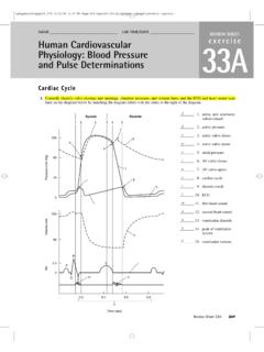
4 2 8. Measuring Heart s Conduction System with EKG > P wave = when atria contract > QRS wave = when ventricles contract > T wave = when ventricles relax > there are normal time intervals between these events. Slower or faster intervals = problem! 9. Heart s Pacemaker Regulation Concept: action potentials spontaneously occur in pacemaker, BUT the RATE of pacemaker depolarization changes. 1. Pacemaker cells depolarize (muscle cells contract) with > opening of Na+ and Ca+2 channels > causes AP or EPSP > cells contract 2. Pacemaker cells repolarize (rest) with > opening of K+ channels > cells relax 10. Regulation of pacemaker depolarization (heart rate) controlled by cardiac center in Medullua > Sympathetic innervation of heart through Thoracic & Cardiac nerves (Review of Ch 6 & 4) > ASK: What is the neurotransmitter for sympathetic change in HR?
5 ANS = epinephrine. > ASK: What is the receptor for epinephrine on the heart? ANS = 1 adrenergic > ASK: What ion channels are opened? ANS = Na+ and Ca+2 channels > ASK: What does that do to HR? ANS = increased > Parasympathetic innervation of heart through Vagus nerves > ASK: What is the neurotransmitter for parasympathetic? ANS = Acetylcholine (ACh) > ASK: What is the receptor on heart for ACh? ANS = muscarinic cholinergic > ASK: What ion channel opened? ANS = K+ channel > ASK: What does that do to HR? ANS = decreases 11. Arrhythmias = abnormal HR (see online clinical app) > Tachycardia = faster than normal HR Treatment: - Na+ channel blockers - Ca+ channel blockers - Beta blockers block 1 adrenergic receptor on heart to slow HR Ex.
6 General beta blocker = propranolol (blocks both 1 & 2) 1 specific blocker = atenolol (blocks only the 1) > Bradycardia = slower than normal HR Treatment: - Digitalis = makes Ca+2 more available and increases strength of heart contraction - 1 Agonist = ASK if they can remember from Ch 4 & 6. ANS = Dobutamine (makes the heart beat faster) - MAO-I class A = ASK if they can remember what this does. ANS = prevents breakdown of epinephrine in synapse with heart muscle. More epinephrine means increased HR. 12. Review slide 13. Blood Pressure = pressure of arterial blood against vessel wall. Can measure with blood pressure cuff. > Systolic BP = pressure from ventricles contracting. Normal range arterial systolic BP = 80 160 mmHg > Diastolic BP = pressure when ventricles relaxed.
7 3 14. Measuring BP with a Sphygmomanometer (they did their first lab experiment of semester doing this. Is REVIEW) 15. What is happening when BP cuff is inflated vs deflating (Korotcoff sounds through stethoscope) 16. Cardiac Output, Cardiac Rate & Stroke Volume Cardiac output = volume of blood ejected by each ventricles per minute Cardiac output = Stroke volume X Cardiac rate (ml/min) (ml/beat) (beats/min) > Average cardiac rate = 70 bpm > Average stroke volume = 70 80 ml/beat > Average cardiac output = 5,500 ml/minute [ L/min] *This is ~ same volume of blood in body! 17. Things that Influence Cardiac Output: 1) End Diastolic Volume (EDV) = the amount of blood in the ventricles during diastole. EDV is affected directly by venous return of blood to heart.
8 As venous return , EDV As venous return , EDV 18. 2) Stretching of the ventricles (as they fill in diastole) Frank Starling (Stretching) Law of the Heart states that: As EDV , ventricle stretch , and so cardiac output As EDV , ventricles stretch , and so cardiac output 19. As stroke volume , so does cardiac output As stroke volume , so does cardiac output Things that affect stroke volume: EDV as EDV , stroke volume , so cardiac output will As EDV , stroke volume , so cardiac output will 4. Heart contractility = strength of heart contraction As contractility , stroke volume , so cardiac output will As contractility , stroke volume , so cardiac output will 5.
9 Total peripheral resistance (TPR) = resistance of blood flow in arteries (depends on blood viscosity, how long vessels are, and their diameter). [imagine blood that is thick like corn syrup, there would be larger resistance of that blood in artery than if blood was less viscous, like water.] As TPR , stroke volume , so cardiac output As TPR , stroke volume , so cardiac output 20. 6. Heart Rate as HR , cardiac output As HR , cardiac output Drugs that cardiac output by HR: A) Epinephrine (through B1-adrenergic receptors) B) Digitalis = makes Ca+1 more available to strength of heart contraction C) MAO-I class A = prevents epinephrine breakdown, causing HR D) Dobutamine = makes da heart beat faster buy acting as B1 adrenergic agonist Drugs that cardiac output by HR: A) General B blockers (propranolol) B) B1 specific blockers (atenolol) C) Na+ channel blockers D) Ca+ channel blockers 4 21.
10 Summary of all things covered that affect cardiac output. ALL of them have direct proportional effect on cardiac output, EXCEPT TPR, which has inverse relationship. 22. 3 ways body regulates cardiac output, blood volume, & blood pressure: 1. Baroreceptor reflex a) Heart baroreceptors (aortic arch & carotid sinus) if BP high ANP release, if BP low inhibit ANP b) Medulla oblongata baroreceptors cardiac & vasomotor center response. Sense low or high BP 2. Hypothalamus ADH release 3. Kidneys - renin-angiotensin-aldosterone system There are practice flow diagrams on the online syllabus for these! Baroreceptors = blood pressure receptors located within aortic arch & carotid sinus. Baroreceptors involved in regulating cardiac output (heart rate) depending on blood pressure in heart.