Transcription of Frog Cardiovascular Physiology - Welcome to Biology!
1 Investigation of human Cardiovascular Physiology is very interesting, but manyareas obviously do not lend themselves to experimentation. It would be tanta-mount to murder to inject a human subject with various drugs to observe theireffects on heart activity or to expose the human heart in order to study the lengthof its refractory period. However, this type of investigation can be done on frogsor computer simulations and provides valuable data because the physiologicalmechanisms in these animals, or programmed into the computer simulation, aresimilar if not identical to those in this exercise, you will conduct the cardiac investigations just Electrical Properties of Cardiac Muscle: Automaticity and RhythmicityCardiac muscle differs from skeletal muscle both functionally and in its fine struc-ture. Skeletal muscle must be electrically stimulated to contract. In contrast, heartmuscle can and does depolarize spontaneously in the absence of external stimula-tion.
2 This property, called automaticity, is due to plasma membranes that have re-duced permeability to potassium ions but still allow sodium ions to slowly leakinto the cells. This leakage causes the muscle cells to slowly depolarize until theaction potential threshold is reached and fast calcium channels open, allowingCa2+entry from the extracellular fluid. Shortly thereafter, contraction spontaneous depolarization-repolarization events occur in a regular andcontinuous manner in cardiac muscle, a property referred to as the following experiment, you will observe these properties of cardiacmuscle in a computer simulation. Additionally, your instructor may demonstratethis procedure using a real Cardiovascular Physiology6 EXERCISEOBJECTIVES1. To list the properties of cardiac muscle as automaticity and rhythmicity,and to define To explain the statement, Cardiac muscle has an intrinsic ability tobeat.
3 3. To compare the relative length of the refractory period of cardiac musclewith that of skeletal muscle, and to explain why it is not possible totetanize cardiac To define extrasystole, and to explain at what point in the cardiac cycle(and on an ECG tracing) an extrasystole can be To describe the effect of the following on heart rate: vagal stimulation,cold, heat, pilocarpine, atropine, epinephrine, digitalis, and potassium,sodium, and calcium To define vagal escape and discuss its To define ectopic 1/10/08 5:23 PM Page 7778 Exercise 6 Nervous Stimulation of the HeartBoth the parasympathetic and sympathetic nervous systems in-nervate the heart. Stimulation of the sympathetic nervous sys-tem increases the rate and force of contraction of the heart. Stim-ulation of the parasympathetic nervous system (vagal nerves)decreases the depolarization rhythm of the sinoatrial node andslows transmission of excitation through the atrio-ventricularnode.
4 If vagal stimulation is excessive, the heart will stop beat-ing. After a short time, the ventricles will begin to beat is referred to as vagal escapeand may be the result of sym-pathetic reflexes or initiation of a rhythm by the Purkinje Frog Heart ActivityThe heart s effectiveness as a pump is dependent both on in-trinsic (within the heart) and extrinsic (external to the heart)controls. In the first experimental series, Activities 1 3, youwill investigate some of these nodal system, in which the pacemaker imposes itsdepolarization rate on the rest of the heart, is one intrinsicfactor that influences the heart s pumping action. If its im-pulses fail to reach the ventricles (as in heart block), the ven-tricles continue to beat but at their own inherent rate, which ismuch slower than that usually imposed on them. Althoughheart contraction does not depend on nerve impulses, its ratecan be modified by extrinsic impulses reaching it through theautonomic nerves.
5 Cardiac activity is also modified by vari-ous chemicals, hormones, ions, and metabolites. The effectsof several of these chemical factors are examined in the nextexperimental series, Activities 4 frog heart has two atria and a single, incompletelydivided ventricle. The pacemaker is located in the sinus veno-sus, an enlarged region between the venae cavae and the rightatrium. The sinoatrial (SA) node of mammals may haveevolved from the sinus Exercise 6: Frog Cardiovascular Physiologyfrom the drop-down menu and click click Electri-cal opening screen will appear in a fewseconds (Figure ). When the program starts, you will see atracing of the frog s heartbeat on the oscilloscope display inthe upper right part of the screen. Because the simulation au-tomatically adjusts itself to your computer s speed, you maynot see the heart tracing appear in real time. If you want toincrease the speed of the tracing (at the expense of tracingquality), click the Tools menu, choose Modify Display, andthen select Increase screen of the Electrical Stimulation 1/10/08 5:23 PM Page 78To familiarize yourself with the equipment, chooseBalloons On/Offfrom the Help menu.
6 This feature allowsyou to scroll around the screen and view equipment can turn off this feature by returning to the Help menuand selecting Balloons oscilloscope display shows the ventricular contractionrate in the Heart Rate window. The heart activity window to theright of the Heart Rate display provides the following messages: Heart Rate Normal displayed when the heart is beatingunder resting conditions. Heart Rate Changing displayed when the heart rate isincreasing or decreasing. Heart Rate Stable displayed when the heart rate issteady, but higher or lower than normal. For example, if youapplied a chemical that increased heart rate to a stable buthigher-than-normal rate, you would see this electrical stimulator is below the oscilloscope display. Inthe experiment, clicking Single Stimulus delivers a singleelectrical shock to the frog heart. Clicking Multiple Stimulusdelivers repeated electrical shocks at the rate indicated in theStimuli/sec window just below the Multiple Stimulus the Multiple Stimulus button is clicked, it changes to aStop Stimulus button that allows you to stop electrical stimu-lation as desired.
7 Clicking the (+)or ( )buttons next to theStimuli/sec window adjusts the stimulus rate. The voltage de-livered when Single Stimulus or Multiple Stimulus is clickedis displayed in the Voltagewindow just below the SingleStimulus button. The simulation automatically adjusts thevoltage for the experiment. The postlike apparatus extendingupward from the electrical stimulator is the electrode holderinto which you will drag-and-drop electrodes from the supplycabinet in the bottom left corner of the left side of the screen contains the apparatus thatsustains the frog heart. The heart has been lifted away fromthe body of the frog by a hook passed through the apex of theheart. Although the frog cannot be seen because it is in thedissection tray, its heart has not been removed from its circu-latory system. A thin string connects the hook in the heart tothe force transducer at the top of the support bracket.
8 As theheart contracts, the string exerts tension on the force trans-ducer, which converts the contraction into the oscilloscopetracing. The slender white strand extending from the heart to-ward the right side of the dissection tray is the vagus nerve. Inthe simulation, room-temperature (23 C) frog Ringer s solu-tion continuously drips onto the heart to keep it moist and re-sponsive so that a regular heart beat is two electrodesyou will use during the experiment arelocated in the supply cabinet beneath the dissection tray. TheDirect Heart Stimulationelectrode is used to stimulate theventricular muscle directly. The Vagus Nerve Stimulationelectrode is used to stimulate the vagus nerve. To position ei-ther electrode, click and drag the electrode to the two-prongedplug in the electrode holder and then release the mouse 1 Recording Baseline Frog Heart beginning to stimulate the frog heart experimen-tally, watch several heartbeats.
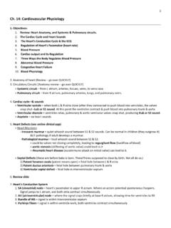
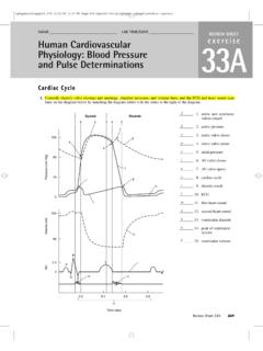
9 Be sure you can distinguishatrial and ventricular contraction (Figure ). the number of ventricular contractions per minutedisplayed in the Heart Rate window under the (beats per minute) ACTIVITY 2 Investigating the Refractory Period of Cardiac MuscleIn Exercise 2 you saw that repeated rapid stimuli could causeskeletal muscle to remain in a contracted state. In otherwords, the muscle could be tetanized. This was possible be-cause of the relatively short refractory period of skeletal mus-cle. In this experiment you will investigate the refractory pe-riod of cardiac muscle and its response to and hold the mouse button on the Direct HeartStimulationelectrode, and drag it to the electrode the mouse button to lock the electrode in electrode will touch the ventricular muscle single shocks in succession by clicking SingleStimulus rapidly. You may need to practice to acquire thecorrect for extrasystoles,which are extra beats that showup riding on the ventricular contraction peak.
10 Also note thecompensatory pause, which allows the heart to get back onschedule after an extrasystole (Figure ).Frog Cardiovascular Physiology79 One-second time line(a)One-second time line(b)AtrialsystoleVentricular systoleVentricular diastoleNormal systoleExtrasystoleCompensatorypauseFIGU RE of contractile activity of a frog heart.(a) Normal heartbeat. (b) Induction of 1/10/08 5:23 PM Page 7980 Exercise 6On the basis of the recording, during which portion of thecardiac cycle was it possible to induce an extrasystole? UseFigures and b to help you to tetanize the heart by clicking Multiple Stim-ulus. Electrical shocks will be delivered to the muscle at arate of 20 stimuli/sec. What is the result?Considering the function of the heart, why is it important thatthe heart muscle cannot be tetanized? Stop Stimulus to stop the electrical stimulation. ACTIVITY 3 Examining the Effect of Vagus Nerve StimulationThe vagus nerve carries parasympathetic impulses to theheart, which modify heart the Direct Heart Stimulationelectrode to returnit to the supply and drag the Vagus Nerve Stimulationelectrodeto the electrode the mouse button to lock the electrode in vagus nerve will automatically be draped over the elec-trode the stimulator to 50 stimuli/sec by clicking the(+)or ( ) Multiple Stimulus.