Transcription of Cardiovascular Physiology - Jones & Bartlett Learning
1 Sebastian Kaulitzki/ShutterStock, PhysiologyCase 1 IntroductionThe Anatomy of the Cardiovascular System: The HeartCardiac Muscle: Cellular Level of OrganizationAnatomy: The VasculatureNutrient ExchangeHow Does Blood Return to the Heart?Blood: What Is Flowing Through These Vessels?What Causes the Heart to Beat?What Is an ECG and What Information Does It Provide?What Is Cardiac Output and How Is It Regulated?Metabolism, O2 Consumption, and Cardiac WorkBlood Vessels Carry Blood to Tissues The End UsersBlood Flow: Behavior of FluidsSummaryKey ConceptsKey TermsApplication: PharmacologyCardiovascular Clinical Case: Type 2 Diabetes 14311/4/2013 8:03:32 PM Jones & Bartlett Learning , LLC.
2 NOT FOR SALE OR DISTRIBUTION144 CHAPTER 7 Cardiovascular PhysiologyIntroductionThe Cardiovascular system consists of the heart and the connecting vasculature, from aorta to arterioles to capillaries to veins to vena cavae. It functions as the distributor of molecules to the billions of cells in the body. Hormones are transported to their target cells via the blood. Nutrients absorbed during digestion are delivered to cells via the circulation, and some of the waste products of cells go to the kidney for elimination, carried in the blood supply. Oxygen (O2), essential for adenosine triphosphate (ATP) production, is carried in the blood, as is the gaseous metabolic waste product, carbon dioxide (CO2).
3 Transportation of all of these molecules is dependent upon the constant movement of blood within the cir-culatory system. This movement is achieved through the actions of a pump, our heart, and a series of non-rigid, living pipes, the Anatomy of the Cardiovascular System: The HeartThe heart is a muscular organ, approximately the size of your fist, comprising four chambers: left and right atrium and left and right ventricles. It is composed of striated muscle, similar to skeletal muscle, but instead of contracting against fixed attachments to bone, as skeletal mus-cle does, it contracts against an incompressible fluid, blood. So the heart contracts against a hydrostatic skeleton.
4 The walls of the atria are thin, reflecting the low pressure exerted by blood returning to the heart. The atria connect to their respective ventricles by atrioven-tricular (A-V) valves, which open whenever the pressure in the atrium exceeds that of the ventricle. These valves are in turn connected to the ventricular wall by papillary muscles and chordae tendineae, lengths of connective tissue that gave rise to the name heartstrings (FIGURE ). These muscles contract whenever the ventricles contract, keeping the A-V valves closed during ventricular contraction. This prevents blood from returning to the right ventricle is a thin-walled chamber, as it only needs to exert enough pressure to force blood from the ventricle to the nearby lung.
5 The conducting vessels of the lung quickly divide into a vast capillary bed, so there is little resistance to the flow of blood. The left ventricle, however, must generate enough force to pump blood to the entire systemic circulation. To achieve this, the left ventricle is a thick-walled chamber that contracts in a spiral fashion (remember that the heart resembles a cone in shape) with sufficient force to open the semilunar valve of the aorta and pump blood throughout the body. Like skeletal muscle, cardiac muscle can hypertrophy after repeated bouts of heavy use, increasing the thickness of the ventricular wall and improving its ability to generate forceful contractions.
6 This means your morning exercise increases your heart strength as well as your leg strength!The heart serves as the pump of the Cardiovascular system, providing the pressure head to move fluid, blood. As mentioned earlier, the heart is four-chambered, but it is best thought Case 1 You generally go for a 3-mile run in the morning, and today is no exception. Within the first minute, you feel your heart rate increase, and before long you can feel the pounding of your heart. As your run continues, you think about the blood coursing through your arteries and veins and wonder at the workings of the Cardiovascular system. What caused your heart rate to increase?
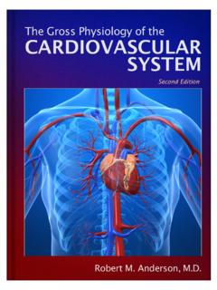
7 What makes your heart beat harder? What makes you flush by the end of the run? How is all this regulated, so that it returns to normal at the end of your run? 14411/4/2013 8:03:32 PM Jones & Bartlett Learning , LLC. NOT FOR SALE OR DISTRIBUTIONThe Anatomy of the Cardiovascular System: The Heart145of as two separate pumps functioning in parallel, with each atrium filling simultaneously, draining into the ventricles simultaneously and the ventricles contracting simultaneously. The right atrium receives blood from the superior and inferior vena cavae. Blood moves passively into the right ventricle following a simple pressure gradient. Contraction of the right ventricle AortaAortaLeft pulmonary arteriesPulmonarytrunkPulmonary semilunarvalveAortic semilunar valvePulmonary veinsPulmonary veinsChordae tendineaeLeft AtriumRight AtriumPapillary musclesAnterior cardiac veinMarginal branchSmall cardiac veinRight atrioventricular (tricuspid valve)Left atrioventricular (bicuspid valve)Left coronary arteryRight coronary arteryCircumflex branchAnterior interventricular branchGreat cardiac veinLeft VentricleRight VentricleInferior Vena CavaSuperior Vena CavaFIGURE The structure of the heart and the path of blood flow through 14511/4/2013 8:03:40 PM Jones & Bartlett Learning , LLC.
8 NOT FOR SALE OR DISTRIBUTION146 CHAPTER 7 Cardiovascular Physiologypumps blood into the pulmonary artery and then into the nearby pulmonary circulation of the lung. There, blood is intimately exposed to air in the alveoli, releasing CO2 and binding O2. Pulmonary veins collect this oxygenated blood and return it to the left atrium through the pulmonary veins. Again, the blood moves passively, down a pressure gradient, through the A-V valve, into the left ventricle. Upon contracture, the left ventricle pumps blood through the semilunar valve into the aorta, for its transit through the vast systemic circulation. Blood will ultimately reach each cell of the body, delivering nutrients, hormones, and O2 while pick-ing up CO2, metabolic waste products, and metabolites before being returned to the right atrium via the vena cava (Figure ).
9 Cardiac Muscle: Cellular Level of OrganizationAt the tissue level, cardiac muscle resembles skeletal muscle, in that it contains regular arrays of thick filaments (myosin) and thin filaments (actin and associated regulatory proteins) arranged within sarcomeres, bounded by Z disks. During contraction, cardiac muscle works on the same sliding filament mechanism described in skeletal muscle. However, there are important differences. Cardiac muscle fibers contain only one nucleus, instead of the mul-tiple nuclei of skeletal muscle. Cardiac muscle fibers branch instead of being consistently linear. Most importantly, cardiac muscle fibers are connected to one another by intercalated disks, which contain gap junctional proteins.
10 These proteins are most abundant on the longitudinal axis of the muscle cells and allow very low resistance conduction between two cardiac muscle cells. It is analogous to adjacent hotel rooms joined by a connecting door transit between the two is extremely fast, becoming essentially a single room. Gap junc-tional proteins allow the heart muscle to function as a syncytium, that is, as though heart muscle cells were one large muscle fiber, instead of thousands of individual mus-cle cells (FIGURE ). Simultaneous contraction is essential if coordinated force is to be generated to pump blood out of the heart on each beat.