Example: biology
MITRAL VALVE STENOSIS - asecho.org
Depends on cardiac output 4. 1.5 cm2 PHT = 29% of total deceleration time (DT) MVA = 220 / Pressure half time MVA = 750 / Deceleration time Deceleration time. 11 ... • Mitral valve area measurement – > 1.5 cm2-Mild – 1.1 to 1.5 cm2-Moderate – < or = 1.0 cm2-Severe . Title: Microsoft PowerPoint - 0506-1000-Levine-Mitral Stenosis ...
Tags:
Information
Domain:
Source:
Link to this page:
Documents from same domain

Guidelines for Cardiac Sonographer Education ...
asecho.orgcardiac sonographer. Specific goals of this revision include the following: • Define the role of the cardiac sonographer • Provide recommendations for educational path-

Aortic Regurgitation: Etiology and Echo …
asecho.orgAortic Regurgitation: Etiology and Echo Quantification Martin G. Keane, MD, FASE Professor of Medicine Lewis Katz School of Medicine at Temple University

Recommendations for Noninvasive Evaluation of …
asecho.orgASE GUIDELINES AND STANDARDS Recommendations for Noninvasive Evaluation of Native Valvular Regurgitation A Report from the American Society of Echocardiography
Recommendations for the Evaluation of Left …
asecho.orgASE/EACVI GUIDELINES AND STANDARDS Recommendations for the Evaluation of Left Ventricular Diastolic Function by Echocardiography: …

Echocardiography in Pulmonary Arterial …
asecho.orgSTATE-OF-THE-ART REVIEWARTICLES Echocardiography in Pulmonary Arterial Hypertension: from Diagnosis to Prognosis Eduardo Bossone, MD, PhD, Antonello D’Andrea, MD, PhD, Michele D’Alto, MD, Rodolfo Citro, MD,
Recommendations for Evaluation of Prosthetic …
asecho.orgGUIDELINES AND STANDARDS Recommendations for Evaluation of Prosthetic Valves With Echocardiography and Doppler Ultrasound A Report From the American Society of Echocardiography’s Guidelines and Standards

2017 Coding and Reimbursement Newsletter. …
asecho.org{D0700524.DOCX / 1 } o If you perform moderate sedation in conjunction with the TEE or other procedures that you provide, use the following new CPT
Left Atrial Appendage Closure Devices - asecho.org
asecho.org10/10/2017 1 Muhamed Sarić MD, PhD, MPA Director of Noninvasive Cardiology | Echo Lab Associate Professor of Medicine Left Atrial Appendage Closure Devices

Multimodality Imaging Guidelines of Patients with ...
asecho.orgASE GUIDELINES AND STANDARDS Multimodality Imaging Guidelines of Patients with Transposition of the Great Arteries: A Report from the American Society of Echocardiography Developed

EAE/ASE Recommendations for Image Acquisition …
asecho.orgTABLE OF CONTENTS 1. Introduction 4 2. Instrumentation 4 a. Fully Sampled Matrix-Array Transducers 4 3. Data Acquisition 5 a. Challenges with 3DE Ac-
Related documents
Coronary Diagnostic and Intervention Coding Sheet - Medtronic
asiapac.medtronic.comcardiac output measurement (separate procedure) 93561 Indicator dilution studies, subsequent measurement of cardiac output 93562 Intravascular Doppler velocity and/or pressure-derived coronary flow reserve +93571 Intravascular Doppler …
Basic Cardiac Rhythms Identification and Response
utmc.utoledo.eduCardiac output (CO) Amount of blood pumped by the left ventricle in one minute Normal cardiac output is 4 – 8 L/min Cardiac Output = ... measurement of: Time (rate) Measured on the horizontal line Amplitude (voltage) Measured on the vertical line .
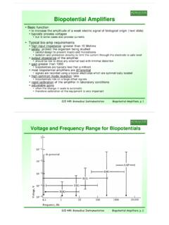
Biopotential Amplifiers - Michigan State University
www.egr.msu.edu• output impedance of the amplifier ... • dipole represented as a cardiac vector M • Mi th di l tM is the dipole moment ... source of the electric field and the measurement systemsource of the electric field and the measurement system • very important for EEG measurement
The Gross Physiology of the Cardiovascular System
cardiac-output.infoAppendix: Clinical Measurement of the Mean Cardiovascular Pressure 61 About the Author 63 Additional Resources 64 . The Gross Physiology of the Cardiovascular System │ 1 Introduction At a time when knowledge about microvascular physiology and subcellular ... increase cardiac output.

DETAILED STATISTICAL ANALYSIS PLAN (SAP) - ClinicalTrials.gov
clinicaltrials.govvariables urine output and capillary refill time an example (7). In our design paper, the SD of cardiac index was 0.99 and the SD of urine output was 0.98, with a slope estimate of 0.040 obtained when cardiac index was regressed against urine output. The SD of capillary refilltime was 2.4, with a slope
ISUOG Practice Guidelines: performance of firsttrimester ...
www.isuog.orgconfirm the presence of an embryo with cardiac activity at the time of examination. Embryonic cardiac activity has been documented in normal pregnancies at as early as 37 days of gestation29, which is when the embryonic heart tube starts to beat30. Cardiac activity is often evident when the embryo measures 2 mm or more31, but is not

Respiratory Therapy Normal Values
www.respiratorytherapyzone.comC. Cardiac Index of 3.7 L/min/m2 D. CVP of 5 cmH2O To get this one correct, you simply needed to know the normal values of the hemodynamic data that is listed in the answer choices. If you can interpret that, you can easily come up with the correct answer. • Normal shunt is 5% or less. • Normal SVR is 1400 dyn/s/cm-5