Transcription of Part II: Sample Preparation for AFM Particle Characterization
1 Part II: Sample Preparation for AFM Particle Characterization Natasha Starostina, Paul West Pacific Nanotechnology, Inc. 3350 Scott Blvd., Suite 29. Santa Clara, CA 95054. Part II: Sample Preparation for AFM Particle Characterization 1. Natasha Starostina, Paul West Pacific Nanotechnology 3350 Scott Blvd. Santa Clara, CA 95054. Abstract Scanning Probe Microscopy has been routinely employed as a surface Characterization technique for nearly 20 years. Atomic Force Microscopy is the most widely used subset of SPM, which can be used in ambient conditions with minimum Sample Preparation . AFM is able to measure three-dimensional topography information from the angstrom level to the micron scale with unprecedented resolution. This paper reviews the most common methods of Sample Preparation that are used for imaging nanoparticles with an AFM. AFM is well suited to individual Particle Characterization . The standard set of measured parameters includes: volume, height, size, shape, aspect ratio and Particle surface morphology.
2 As a single- Particle technique, physical parameters for each Particle in an image can be recorded and the data set can be processed to generate a statistical distribution for an entire set of particles ( ensemble-like information). Speeding up the process of obtaining data is critical for many reasons and de nitely makes AFM more attractive given its ability of individual Particle imaging. In general, the AFM Particle Characterization is both cost and time effective as well as easier to use than electron microscopy. The resolution of AFM is greater or comparable to that of SEM/TEM, and strong advantages of AFM for Particle Characterization include direct measurements of height, volume and 3D display. Introduction Over the past 20 years Scanning Probe Microscopes (SPM) have emerged as an essential material Characterization technique in various fields1,2,3,4,5. The importance of the SPM was evident as early as 1984 when the Nobel prize was awarded for the Scanning Tunneling Microscope (STM) invention by IBM researchers1.
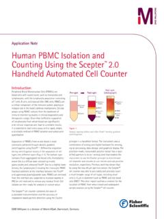
3 Today the Atomic Force Microscope (AFM) is the most commonly used scanning probe technique for materials characterization2. Major advantages of AFM are that it has a combination of high resolution in three dimensions, the Sample does not have to be conductive, and there is no requirement for operation within a vacuum. With an AFM, a large range of topographies and many types of materials can be imaged. Examples of surface features that may be imaged include: atomic terraces, carbon nanotubes, colloidal particles , viruses, DVD textures up to micro lens textures, fractured surfaces, and complex multi-phase polymers. In other words, AFM is capable of delivering unique 3D topography information from the angstrom level to the micron scale with unprecedented resolution. With an AFM, the Z-axis resolution ( , perpendicular to the Photo Detector Laser surface) is typically better than the resolution in the XY scan plane of the Sample surface.
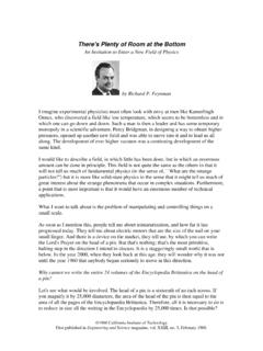
4 Under ambient conditions, the Z- So resolution for most of the commercially available AFMs is on the Cantilever Differential Amplifier sub-angstrom level. Resolution in the X-Y scan place is oftern limited by the diameter of the probe and is on the order of a few nanometers. In the X and Y ases, AFM images are always a Sample convolution of the probe geometry and Sample texture. However, if the probe is much smaller than the surface features, the image Figure 1: Schematic of AFM. distortions introduced by the probe are minimal. The extreme sensitivity of the AFM is derived from a force sensor that measures forces between the probe and target surface which are typically less than 1 nN/nm. Most AFM's utilize a light lever or a force sensor, as first disclosed in 1929 and then applied to the AFM in 1986. Figure 1 shows a schematic of the AFM. Recently, a new type of force sensor, based on a crystal resonator, shows promise for making the AFM much simpler to operate6,7 and it provides a very high force sensitivity required for high resolution imaging.
5 Part II: Sample Preparation for AFM Particle Characterization 2. Table 1: Given the wide variety of applications that use particles , it makes sense that there are many different ways to analyze and characterize particles . The following is a partial list of the material classes of particles in different environmental media, as analyized by an AFM: Imaging in Air Imaging in liquids Imaging embedded particles Dry Powders Bio- particles in Buffer Soft polymer and Bio-materials Evaporated Suspensions Inorganic particles Hard Surface Materials Bio- particles Membranes and defects Carbon nanotubes TEM samples SEM samples For Particle Characterization , there is no single instrument that Liquid-borne methods Air-borne methods is the right tool for every job . In fact, more than 400 different techniques exist for Particle counting, sizing, analyzing, and characterizing. Typically, instrumentation is chosen by engineers and researchers through consideration of what measurements Ensemble need to be made, and in what environment the measurements methods need to be made.
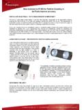
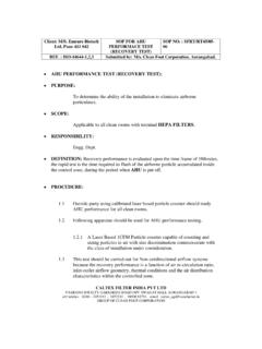
6 There are two primary considerations for AFM. selecting instrumentation: a) single Particle versus ensemble, and b) the environment the measurement is made in air, TEM. liquid, vacuum. Table 1 shows a comparison of the most SEM Single- Particle methods common material classifications and environmental media for Particle analysis. In general, morphological information, such Figure 2: Particle analysis can vary depending on the air or liquid borne or as shape and aspect ratio, as well as surface information, such ensemble or single- Particle . as texture and roughness parameters, cannot be obtained using ensemble techniques. As presented in Figure 2, there are three methods of characterizing nanoscale particles -- SEM, TEM, and AFM . TEM. TEM and SEM are examples where an established single- Particle Characterization technique is combined with image Time SEM. processing to measure and analyze particles . The emergence Optics AFM. of the application of AFM to nanoparticle Characterization has developed over the past 10 years.
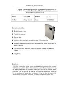
7 AFM is well suited to individual Particle Characterization including: parameters, volume, size, shape, and Particle surface morphology. With Cost single- Particle techniques, physical parameters for each Figure 3: Analysis of the time and cost for instrumentation of microscope techniques used for characterizing particles . Particle in a set of particles can be recorded and the data set can be processed to generate a statistical distribution ( ensemble-like information) for the entire set of particles . A major factor that contributes to microscope single Particle analysis (and ultimately the accuracy of measurements) is image quality. The AFM is similar to electron microscope techniques, SEM and TEM, where proper Sample Preparation is the key to measuring high quality data. The art of Sample Preparation is in fact a simple procedure of critical-path steps, where every single step makes a difference. TEM is well known for very time-consuming and complicated Sample preparation8,9.
8 SEM samples are easier to prepare, however the requirement for conductivity adds some difficulty. AFM samples do not have to be conductive, which makes Sample Preparation easier for the user, Figure 3. However, there are very important criteria to be met in order to do AFM imaging. Part II: Sample Preparation for AFM Particle Characterization 3. The main focus of this article is to present a general survey of AFM Sample Preparation methods for nanoparticle Characterization . The authors present a review of the available methods for AFM Particle imaging and Characterization in ambient conditions and in liquids. When available, references for additional information are provided. All AFM images shown is this paper have been obtained on a Light Lever or Crystal Nano-RP AFM. Close contact mode is the imaging mode for all images scanned in air this paper. particles , Dispersion, Substrates, Adhesives The vast variety of particles can be categorized as engineered and non-engineered environmental particles .
9 Engineered or artificially created particles break down to organic and inorganic categories. Those categories in turn have additional subdivisions, for example powders, suspensions and embedded particles . Different approaches can be used to prepare AFM samples10-16. Particle size, hydrophobicity, native environment and bio-compatibility are taken into consideration. AFM Particle imaging requires that: a) The particles to be rigidly adhered to a substrate. b) The particles to be dispersed on a the substrate. c) The substrate roughness is less than the size of the nanoparticles . Often an adhesive is required for affixing the nanoparticles on a substrate. There are a large number of adhesive choices of the adhesive for small Particle deposition. The most commonly used chemicals are poly-l-lysine, poly-D-lysine17, PEI (poly-ethyleneimide) or APTES (aminopropyltriethoxy silane) to facilitate chemical bonding between Particle and substrate.
10 Functionalized surfaces might either promote adsorption or allow covalent bonding. Sometimes hydrophobic substrates are preferable in air-AFM, this way formation of a water monolayer can be avoided12. HPOG or spin-coating of polymers is a convenient method for creating hydrophobic substrates. If imaging needs to be done in an aqua-solution then hydrophilic agents should be used. Particle -substrate affinity has to be stronger than tip to Particle interaction. Buffer solution chemical composition, pH, can be modified to maximize the adhesion between particles and the substrate, when imaging in liquid10. If the nanoparticles are not dispersed on the substrate it is not possible to characterize them. Establishing the optimal method for dispersing nanoparticles on a substrate often requires experimentation. The challenge of dispersing nanoparticles on the substrate is great primarily because of several competing factors. On a large scale, exposure time and dilution of the Particle solution must be considered.