Transcription of Rehabilitation Guidelines for Elbow Ulnar Collateral ...
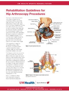
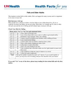
1 UW HEALTH SPORTS REHABILITATIONThe world class health care team for the UW Badgers and proud sponsor of UW 621 SCIENCE DRIVE MADISON, WI 53711 4602 EASTPARK BLVD. MADISON, WI 53718 Rehabilitation Guidelines for Elbow Ulnar Collateral Ligament (UCL) ReconstructionThe Elbow is a complex system of three joints formed from three bones; the humerus (the upper arm bone), the ulna (the larger bone of the forearm, on the small finger side) and the radius (the smaller bone of the forearm on the thumb side). This complex system allows a hinging action (bending and straightening) and a rotation action. The stability of the Elbow joint is maintained by the bony congruency, the muscular attachments and the are several important ligaments in the Elbow .
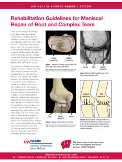
2 Ligaments are soft tissue structures that connect bones to bones. The ligaments around a joint usually combine together to form a joint capsule. A joint capsule is a watertight sac that surrounds a joint and contains lubricating fluid called synovial fluid. In the Elbow , two of the most important ligaments are the Ulnar Collateral ligament (UCL) and the lateral Collateral ligament (LCL). The UCL is also known as the medial Collateral ligament. The UCL is on the medial (the side of the Elbow that s next to the body when your arms are at your side with your palms up or facing out in front of you) side of the Elbow and LCL is on the outside of your Elbow . The Ulnar Collateral ligament is a thick band of tissue that forms a triangular shape along the inside of the Elbow .
3 It has an anterior bundle, posterior bundle and a thinner, transverse ligament. These ligaments can be torn when there is an injury or dislocation of the Elbow . If the injury to the ligament(s) affects the stability of the joint, it is possible that the function of the Elbow will be to the UCL in overhead athletes has been widely reported. Normal activities of daily living rarely place enough stress on the UCL to create instability but throwing sports place high stresses on the Elbow supporting structures. Over time, the high repetitive stresses associated with throwing and overhead activity may create overload to the supporting ligamentous support, resulting in a UCL tear.
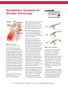
4 Typically, athletes with UCL injury report a history of repetitive throwing with complaints of pain at the medial (inside) aspect of the Elbow during or after their activity. Onset occurs from either one traumatic incident or can develop throughout a long period of time due to repetitive Elbow stress. Eventually the athlete loses their velocity and accuracy of throwing. More than 40% of athletes with UCL injury also report symptoms of Ulnar nerve irritation from friction or snapping of the nerve during overhead thrower often experiences pain with the arm fully cocked (shoulder in full external rotation or the arm rotated all the way back) and as it accelerates through the throw and release of the ball.
5 While throwing, the Elbow can straighten at speeds of over 2300 degrees per second and may have a valgus (side) force that exceeds the ultimate strength of the normal uninjured UCL. Proper mechanics and optimal strength and endurance of the muscles of the upper extremity are needed to assist with injury prevention. Trauma or injury to the UCL results in significant functional limitations including medial Elbow pain, loss of velocity and accuracy with throwing, instability, neurologic (nerve) symptoms and decreased muscular consequences of this injury usually leave the athlete who has a torn UCL with two options: Rehabilitation with activity modification ( avoidance of pitching and performance throwing) or surgical reconstruction with postoperative Rehabilitation prior to return to pitching and performance reconstruction surgery is performed through an incision on the medial (inside) side of the Elbow joint.
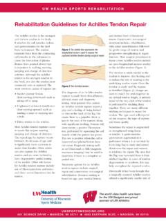
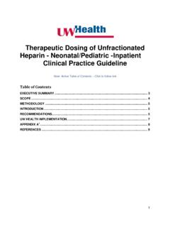
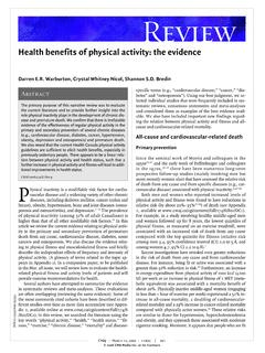
6 The damaged Ulnar Collateral ligament is replaced with a tendon taken from somewhere else in the body. The tendon graft can come from the patient s own forearm, hamstring, knee or foot. This is called an autograft. This tendon is weaved through drill holes in the humerus and ulna to re-create the Rehabilitation Guidelines for Elbow Ulnar Collateral Ligament (UCL) 621 SCIENCE DRIVE MADISON, WI 53711 4602 EASTPARK BLVD. MADISON, WI 53718triangular shape of the common technique used to replace the damaged Ulnar Collateral ligament is called the docking technique. The surgeon drills two holes in the ulna and three in the medial epicondyle of the humerus (the small bump of bone on the inside of the Elbow at the bottom of the upper arm).
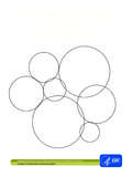
7 The two holes in the ulna form a tunnel that the tendon graft will be looped through. The three holes in the medial epicondyle form a triangle. The bottom hole will be bigger than the top two holes, so that the surgeon can slide the end of the tendon graft into the bottom hole. The two top holes are used to pull the tendon graft into the tunnel using sutures that are attached to the graft and threaded through the two holes. After the tendon is harvested, sutures are attached to both ends. The tendon is looped through the lower tunnel formed in the ulna and stretched across the Elbow joint. The two sutures attached to the ends of the graft are threaded into the larger bottom tunnel in the medial epicondyle and each is threaded out one of the upper, smaller holes.
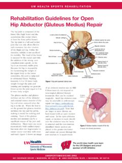
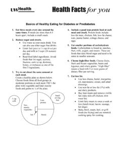
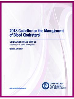
8 Using these two sutures, the surgeon pulls the end of the graft farther into the upper tunnel until the amount of tension is correct to hold the joint in position. The surgeon carefully puts the Elbow through its full arc of motion and readjusts the tension on the sutures until satisfied that the proper ligamentous tension is restored. The two sutures are tied together to hold the tendon graft in that common technique to reconstruct the UCL is called the figure of eight technique. In this technique, the tendon graft is threaded through two pairs of holes - two drilled in the medial epicondye of the humerus and two in the ulna. The graft is looped through the holes in a figure of eight fashion.
9 The two ends of the tendon are sutured to the tendon the muscles on the inside of the Elbow joint and forearm (the flexor muscles of the wrist) were completely detached from the humerus. Now, the flexor muscles are not detached, but are split and retracted to allow the surgeon to see the areas of the Elbow joint required to perform the operation successfully. If there is any concern that the Ulnar nerve has been stretched and damaged due to the instability (as mentioned above), it may be re-routed so that it runs in front of the Elbow joint rather than through the cubital tunnel in the back of the Elbow . The incision is sutured together and the Elbow is placed in a large bandage and following surgical reconstruction of the UCL begins with range of motion and initial protection of the reconstruction, along with resistive exercises to keep the shoulder and core strong.
10 This is followed by progressions for resistive exercise that attempt to fully restore strength and muscular endurance to allow for a safe return to throwing and overhead functional activities. These Guidelines also include aerobic training throughout the Rehabilitation process and, for many, a later stage an interval throwing program. This multi-faceted Rehabilitation approach often includes biomechanical video analysis to ensure proper throwing mechanics before an athlete returns to their early phases of post-operative care for UCL reconstructions involve specific time frames, restrictions and precautions to protect healing tissues and the surgical fixation/reconstruction.