Transcription of Reproductive Tract Anatomy and Physiology of the Cow
1 To find more resources for your business, home, or family, visit the College of Agricultural, Consumer and Environmental Sciences on the World Wide Web at Tract Anatomy and Physiology of the CowGuide B-212 Reviewed by Jason Turner1 Cooperative Extension Service College of Agricultural, Consumer and Environmental Sciences 1 Professor/Extension Horse Specialist, Department of Extension Animal Sciences and Natural Resources, New Mexico State the Anatomy and Physiology of the cow s re-productive system is fundamental to good cattle management. Basic knowledge in this area will help producers do a better job of getting cows rebred, especially when using artificial in-semination and estrus synchronization. It will also enable pro-ducers to better understand and control Reproductive diseases and calving ovary is the primary female Reproductive organ and has two important functions: producing the female Reproductive cell (the egg or ovum) and producing the hormones estrogen and progesterone.
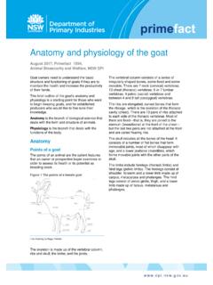
2 The cow s two ovaries are oval to bean-shaped organs that are 1 inches long and located in the abdominal secondary sex organs are a series of tubes that receive semen, transport sperm to the egg so it can be fertilized, nourish the fertilized egg (embryo), and allow the calf to be birthed. These organs include the vagina, cervix, uterus, uter-ine horns, and oviducts (also called Fallopian tubes), which each have a funnel-shaped opening called the infundibulum. Figure 1 presents a diagram of the complete Reproductive Tract ovary produces the egg by a process called oogenesis. In contrast to the continuous production of sperm (spermato-genesis) in the male, oogenesis is cyclic. This cycle (called the estrous cycle) has a characteristic length and consists of a defi-nite sequence of events, both physiological and ovary contains several thousand tiny structures called primary follicles. Each follicle consists of a germ cell sur-rounded by a layer of cells.
3 This germ cell has the potential to mature into an egg if the follicle completes development. However, most of the primary follicles never develop. Rather, they die, are absorbed by the ovary, and are replaced by newly formed primary relatively few primary follicles that develop completely do so through a series of phases. Many layers of cells are added to the single layer of cells surrounding the egg in the primary follicle, forming a central cavity. As the follicle and cavity grow larger, the egg becomes attached to the back of the follicle (opposite the ovulation site) by a stalk of cells. As the follicle continues to grow rapidly, the side opposite the egg bulges from the surface of the ovary and becomes very thin. Once the follicle reaches this mature state it is called a Graafian follicle. At ovulation, the thin portion ruptures to release the contents of the follicle, including the ovulation, the cells that developed within the follicle differentiate to form the corpus luteum, which has the important function of producing released egg is caught by the infundibulum and moves into the oviduct, where fertilization occurs if viable sperm are present.
4 The egg remains capable of fertilization for only a few hours. Thus, it is very important that fertile sperm be pres-ent near the time of ovulation. The egg moves through the oviduct and into the uterus within the next three to four days. If it is fertilized, it begins embryological development in the uterus. If it is not fertilized, it body of the uterus is relatively short and poorly devel-oped, while the uterine horns are longer and well developed. The embryo develops in the uterine horns. The fetus develops within a layer of membranes called the placenta. The mother nourishes the fetus via diffusion of nutrients across the placenta. There is no direct blood connection between fetus and 1. Diagram of the Reproductive Tract of the cow. (Adapted from Rich and Turman, )UterusCervixVaginaVulvaUterine hornsOviductOvaryBladderBlind pouchGuide B-212 Page 2 The cervix is essentially the neck of the uterus. It has thick walls and a small opening that is difficult to penetrate because of overlapping or interlocking folds (annular rings).
5 It serves as a passageway for sperm deposited in the vagina and for the fetus at the time of birth. During pregnancy , it is usually filled with a thick secretion that serves as a plug to protect the uterus from infective material entering through the vagina serves as the receptacle for the male penis dur-ing copulation. During natural mating, semen is deposited in the vagina near the cervix. The urinary bladder opens to the exterior through the urethra, which opens into the va-gina. This region of the vagina is restricted in size because of sphincter muscles associated with the urethral opening. The region behind the external urethral orifice is called the vesti-bule and is a common passageway for both the urinary and the Reproductive systems. The external opening of the vagina is called the REGULATION OF THE FEMALE Reproductive TRACTN ormal reproduction in the female depends on hormones, which are specific chemical substances produced by special-ized glands called endocrine glands.
6 These secretions pass into the blood and lymph fluids and are transported to various parts of the body, where they exert several specific effects. Reproductive hormones may originate in the hypothalamus, pituitary, gonads, uterus, or hormones are produced in the hypo-thalamus, a portion of the brain that is the neural control center for Reproductive hormones. The role of hypothalamic hormones is to regulate the release of other hormones from the pituitary gland, and many hypothalamic hormones are therefore called releasing hormones. The primary releasing hormone of reproduction is gonadotropin-releasing hormone (GnRH). GnRH controls the release of follicle-stimulating hormone (FSH) and luteinizing hormone (LH) from the pi-tuitary gland, located at the base of the brain. These pituitary hormones regulate the production of estrogen and proges-terone from the ovary. FSH stimulates the follicle s growth, development, and function, while LH causes the follicle to rupture and the corpus luteum to develop.
7 The female hormone estrogen is produced by the Graaf-ian follicle on the ovary. Another hormone originating from the ovary is progesterone, which is produced by the corpus luteum. Each has an important role in the female Reproductive regulates several functions: the development and functioning of the secondary sex organs; the onset of heat, or estrus (the period of sexual receptivity); the rate and type of body tissue growth, especially fat deposition; and priming or preparing of the prepubertal heifer and postpartum cow for the onset of sexual is the hormone of pregnancy . It suppresses the further development of follicles and estrogen secretion. While progesterone is being produced, the female does not exhibit estrus. Progesterone is necessary for preparing the uterus to receive the fertilized egg and maintains the proper uterine environment for pregnancy . Estrogen and progester-one are not completely separate in their effects because both are necessary for complete development of some important organs.
8 Uterine development is initiated by estrogen and completed by progesterone. The fertilized egg will not attach and survive in the uterus unless that tissue has been properly prepared by the action of estrogen and progesterone. Estro-gen causes rhythmic contractions of the uterus. On the other hand, progesterone has a quieting effect on the uterus, so there are no contractions that might disturb development of the mammary gland also de-pends on both hormones. Estrogen promotes the growth of the duct system, and progesterone is necessary for the devel-opment of the clusters of milk-secreting alveoli on the ducts. In general, estrogen makes things happen and progesterone calms them number of other hormones and hormone-like chemicals are known to have important roles in regulating the cow s re-productive system. Prostaglandins are secreted by many body tissues and perform a variety of functions. The prostaglandin primarily affecting the cow s estrous cycle is prostaglandin F2 (PGF2 ), which is produced by the uterus.
9 PGF2 is the natural luteolytic agent that causes the corpus luteum to regress late in the estrous cycle and allows a new estrous cycle to be initiated in the nonpregnant female. In a pregnant cow, a signal is sent from the developing embryo to the uterus to prevent PGF2 release, which allows the corpus luteum to persist throughout pregnancy . Maternal recognition of preg-nancy is believed to occur between days 16 and 17 after fertil-ization. Injecting cows or heifers with PGF2 between days 6 and 16 of the estrous cycle will cause premature re-gression of the corpus luteum, with the best results achieved among females injected on days 10 to PGF2 during the first five (1 5) and last five (17 21) days of the estrous cycle will generally not cause luteal regression. The luteolytic response allows the use of PGF2 in estrus synchronization programs in cow herds and initiates abortion in feedlot heifers. Estrogens, progestins, prostaglandins, and GnRH have all been used in various combinations in effective estrus synchronization ESTROUS CYCLEThe cow s Reproductive cycle consists of a series of events that occur in a definite order over a period of days.
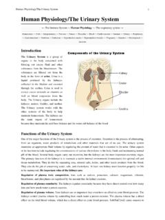
10 The estrous cycle in the cow averages 21 days (range is 17 24). During this time, the Reproductive Tract is prepared for estrus or heat (the period of sexual receptivity) and ovulation (egg release). Figures 2 and 3 outline the sequence of anatomical and hor-monal changes that occur during a typical 21-day cycle in which pregnancy does not B-212 Page 3 Figure 2. Diagram of a 21-day estrous cycle in which pregnancy does not occur. (Adapted from Deutscher, 1980)Day 0: The cow is in estrus (standing heat) due to an increased concentration of estrogen. As estrogen levels reach a certain threshold level, a surge of LH is released by the pituitary. Near the end of standing heat, the mature Graafian follicle ovulates (ruptures) in response to this LH 1 2: The cells that formerly lined the follicle change and become the luteal cells of the corpus luteum. This change in cell form is caused by hormonal ac-tion, primarily the action of 2 5: The corpus luteum grows rap-idly in both size and function.