Transcription of Thoracic aortic aneurysm: Optimal surveillance and …
1 Thoracic aortic aneurysm: Optimal surveillance and treatmentCLEVELAND CLINIC JOURNAL OF MEDICINE VOLUME 87 NUMBER 9 SEPTEMBER 2020 557 Aneurysm of the Thoracic aorta , renal ar- tery, or splenic artery is often detected incidentally but can present acutely with dis-section or rupture, with a high risk of death or morbidities. Computed tomography angiogra-phy (CTA) and magnetic resonance angiogra-phy (MRA) are key to characterizing the an-eurysm and the rest of the vasculature, while ultrasonography or echocardiography assist in assessment and surveillance , and catheter angiography is the gold standard for renal and splenic aneurysm. The need for prophylactic intervention is based on aneurysm size, location, growth, and other associated conditions and risk factors in the individual patient.
2 Management strat-egies include surgery, which is mandatory in the acute setting and in cases of challenging anatomy, and endovascular techniques. Regu-lar imaging surveillance is critical after diag-nosis and after aneurysm interventions. In this, the first of 2 articles, we discuss tho-racic aortic aneurysm (TAA); in the second article, we will discuss renal artery and splenic artery aneurysm. WHAT IS THE CLINICAL IMPORTANCE OF TAA?TAA is clinically important because of the risk of devastating complications acute aortic syn-dromes such as aortic dissection and ,2 Type A aortic dissection (ie, originating in the ascending aorta ) is a fatal condition with dismal in-hospital mortality rates of 57% with-out emergency surgery and 17% to 25% with emergency surgery in national and internation-al registries despite advances in ,4 The mortality rate is much lower but still sig-nificant in expert aortic centers of excellence, of the Thoracic aorta is less common than in the abdominal aorta , but it is clinically important because of the risk of rupture and death.
3 Cases are often found incidentally. Indications for surgical or endovascular repair are based on aneurysm location and risk factors for rupture such as aneurysm size, rate of growth, and associated conditions, while medical management is also important. surveillance with various imaging tests is criti-cal before and after intervention to guide POINTSP atients with bicuspid aortic valve or genetic syndromes such as Marfan syndrome are at higher risk, with lower thresholds for surgical intervention, but account for only a minority of echocardiography has some roles in screen-ing and monitoring the aortic root and ascending aorta , computed tomography and magnetic resonance imaging are necessary for the complete assessment of the Thoracic aorta and are often necessary for from several professional societies are available regarding surveillance and indications for with Thoracic aortic aneurysm require multidis-ciplinary care.
4 Including a cardiologist and possibly a cardiovascular surgeon and genetic counselor. Medical care includes traditional cardiovascular risk fac-tor management. Beta-blockers are often used to control blood pressure but should be used with caution in those with acute aortic valve regurgitation. Tom Kai Ming Wang, MBCHB, MD(res), FRACPA dvanced Cardiac Imaging Fellow, Section of Cardiovascular Imaging, Heart, Vascular, and Thoracic Institute, Cleveland Clinic Milind Y. Desai, MDStaff Cardiologist, Section of Cardiovascular Imaging and Medical Director, aorta Center, Heart, Vascular, and Thoracic Institute, Cleveland Clinic; Professor, Cleveland Clinic Lerner College of Medicine of Case Western Reserve University, Cleveland, OH CMEMOC on February 3, 2022.
5 For personal use only. All other uses require from 558 CLEVELAND CLINIC JOURNAL OF MEDICINE VOLUME 87 NUMBER 9 SEPTEMBER 2020 Thoracic aortic ANEURYSM such as the 4% to 7% reported by Cleveland The incidence of combined TAA and aortic dissection has been reported to be 6 to 13 per 100,000 per year,6 8 although this would underestimate clinically silent There are no effective preventive strategies for TAA to date; thus, early detection, surveil-lance, and treatment are critical to improving outcomes. Guidelines are ,2,9 WHO IS AT RISK?Risk factors for TAA (Table 1) are abundant in modern society and include older age, male sex, hypertension, smoking, and atherosclerosis. No wonder, then, that the incidence of TAA and the number of surgical repairs are ,10 Genetic conditions associated with TAA such as Marfan syndrome are less common but nevertheless important because the prog-nosis and management are ,2,9 Some risk factors or conditions increase wall stress, while others increase medial Although only 5% of cases of TAA are asso-ciated with genetic syndromes, another 20% are in patients who have a family history of TAA, which has important implications for assessment, management, and And many cases are idiopathic, lacking obvi-ous causes or risk factors.
6 HOW IS TAA DISCOVERED?Most cases of TAA are asymptomatic and are discovered either incidentally on imaging or as part of dedicated screening for those at That said, possible symptoms include chest, abdominal, or back pain, dyspnea, cough, dys-phagia, hoarseness, claudication, and cerebro-vascular events. The clinical history should be directed at symptoms, risk factors, and family history. Physical examination should focus on the cardiac, neurologic, and peripheral vascular systems and should include blood pressure (and how it differs in different limbs), pulses, murmurs, and bruits, and other signs specific to associated Basic investigations that can detect pos-sible abnormalities associated with TAA in-clude electrocardiography (showing ischemic changes or myocardial hypertrophy), chest radiography (showing a widened mediastinum or prominent aortic shadow)
7 , and blood tests, including complete blood cell count, metabol-ic profile, and markers of inflammation, coag-ulation, and myocardial injury, many of which help in the differential diagnosis of TAA vs acute aortic ,9 WHAT IS A NORMAL-SIZE aorta ?Although aneurysm is generally defined as an increase of more than 50% of the normal arterial diameter, cardiac imaging guidelines have clear dimension thresholds for different severities of TAA ,10 The risk of rupture or dissection decides who requires prophylactic interventionTABLE 1 Thoracic aortic aneurysm: Risk factors, associations, and causes Risk factors Older age Male sex Hypertension Smoking Hypercholesterolemia Weight-lifting Cocaine use Trauma Cardiovascular associations Atherosclerosis Bicuspid aortic valve Other aneurysm Prior aortic dissection aortic coarctationGenetic causes Familial Thoracic aortic aneurysm Marfan syndrome Loeys-Dietz syndrome Ehlers-Danlos syndrome Turner syndrome Autosomal-dominant polycystic kidney disease Shprintzen-Goldberg (craniosynostosis) syndromeInflammatory causes Takayasu arteritis Giant-cell arteritis Beh et arteritis Ankylosing spondylitisInfective causes Mycotic aortitis SyphilisIdiopathic on February 3, 2022.
8 For personal use only. All other uses require from CLEVELAND CLINIC JOURNAL OF MEDICINE VOLUME 87 NUMBER 9 SEPTEMBER 2020 559 WANG AND DESAI The aorta is larger in men and in larger people generally, and therefore sex and body size are taken into account when determining the normal ranges and severity The aorta also tends to increase in size with age. The upper limit of normal for aortic di-mensions is 2 standard deviations above the mean diameter in a population of healthy adults. aortic dimensions are measured at right angles to the direction of blood flow. On echo-cardiography, the standardized aortic measure-ments are taken in the end-diastolic frame and from leading edge to leading edge for repro-ducibility.
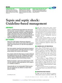
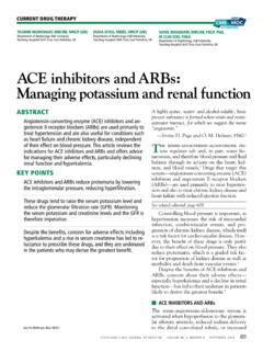
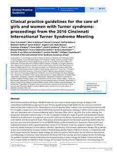
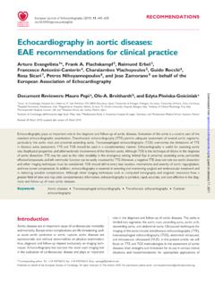
9 On CTA and MRA, measurements are from inner edge to inner edge, from aor-tic sinus to sinus, or from sinus to commissure (often about 2 mm smaller than from sinus to sinus; Figure 1).12,13 The full Thoracic aor-tic study should include measurement of all segments: aortic sinus; sinotubular junction; proximal, mid, and distal ascending aorta ; aor-tic arch; and descending aorta , as well as the maximal dimensions, branch involvement, and surgical The aortic walls should be examined for calcification, throm-bus, dissection, hematoma, and infection. WHAT IMAGING MODALITIES ARE USED? aortic imaging remains central to TAA diag-nosis and ,2,9 Three-dimensional multiplanar recon-struction software for CTA and MRA has rev-olutionized measurement of the aorta , recon-3-D CTA and MRA have revolutionized measurement of the aortaFigure 1.
10 Computed tomography angiography aortic root measurements on (A) axial source image and (B D) 3-dimensional multiplanar reconstruction (3-D-MPR) double-oblique planes. Note that without 3-D-MPR, the aortic root size is underestimated (A). Also note that sinus-commissure measurements are often slightly less than sinus-sinus measurements in (B). on February 3, 2022. For personal use only. All other uses require from 560 CLEVELAND CLINIC JOURNAL OF MEDICINE VOLUME 87 NUMBER 9 SEPTEMBER 2020 Thoracic aortic ANEURYSM structing source images into double-oblique planes to ensure measurements are taken per-pendicular to the lumen (Figure 1).1,2,9 Echocardiographic aortic root measure-ment has the strongest evidence base for guid-ing intervention, and its thresholds have been extrapolated to other modalities and aortic lo-cations.