Transcription of THROMBOELASTOGRAPHY (TEG) IN TRAUMA
1 DISCLAIMER: These guidelines were prepared by the Department of Surgical Education, Orlando Regional Medical Center. They are intended to serve as a general statement regarding appropriate patient care practices based upon the available medical literature and clinical expertise at the time of development. They should not be considered to be accepted protocol or policy, nor are intended to replace clinical judgment or dictate care of individual patients. THROMBOELASTOGRAPHY (TEG) IN TRAUMA . SUMMARY. THROMBOELASTOGRAPHY (TEG) is a test of whole blood coagulation that was developed in the 1950's, but was largely passed over in favor of conventional coagulation tests (PT, PTT, platelet count).
2 It has been applied to transplant and cardiac surgery since the 1980's and has been shown to decrease usage of blood products and mortality when used to evaluate the coagulation cascade during these procedures. With new understanding of the coagulopathy of TRAUMA , there has been considerable interest in the use of TEG in the TRAUMA population. This guideline reviews the interpretation of a thromboelastogram and how it may be used to guide blood product administration. RECOMMENDATIONS. Level 1. None . Level 2. THROMBOELASTOGRAPHY may be used to screen patients for coagulopathy in the following situations: Blunt or penetrating TRAUMA patients who arrive in hemorrhagic shock Patients receiving massive transfusion protocol (1:1:1) to evaluate for discontinuation or guided product therapy Clinical suspicion for hemorrhage or coagulopathy Level 3.
3 THROMBOELASTOGRAPHY may be used to guide blood product administration in bleeding patients as follows: TEG-ACT > 140 or R-time > 10 transfuse fresh frozen plasma K-time > 3 or angle < 53 transfuse cryoprecipitate MA < 50 transfuse platelets LY30 > 3% administer tranexamic acid INTRODUCTION. TEG was developed in 1948 by Hellmut Hartert in Heidelberg, Germany as a test to detect clotting factor deficiencies (1). Its availability in the United States was limited until the 1980's, when the addition of disposable components, tissue activators, and computerized software made the test more practical, reproducible, and timely.
4 A TEG is designed to assist the clinician in identifying whether a patient has normal hemostasis or is bleeding due to a coagulopathy or anticoagulant therapy. If a coagulopathy is identified, the TEG results will point to the specific therapy to treat it, whether using fresh frozen plasma, cryoprecipitate, or an anti-fibrinolytic or thrombolytic drug. EVIDENCE DEFINITIONS. Class I: Prospective randomized controlled trial. Class II: Prospective clinical study or retrospective analysis of reliable data. Includes observational, cohort, prevalence, or case control studies.
5 Class III: Retrospective study. Includes database or registry reviews, large series of case reports, expert opinion. Technology assessment: A technology study which does not lend itself to classification in the above-mentioned format. Devices are evaluated in terms of their accuracy, reliability, therapeutic potential, or cost effectiveness. LEVEL OF RECOMMENDATION DEFINITIONS. Level 1: Convincingly justifiable based on available scientific information alone. Usually based on Class I data or strong Class II. evidence if randomized testing is inappropriate. Conversely, low quality or contradictory Class I data may be insufficient to support a Level I recommendation.
6 Level 2: Reasonably justifiable based on available scientific evidence and strongly supported by expert opinion. Usually supported by Class II data or a preponderance of Class III evidence. Level 3: Supported by available data, but scientific evidence is lacking. Generally supported by Class III data. Useful for educational purposes and in guiding future clinical research. 1 Approved 12/04/2013. Revised 12/3/2014. Specimen handling is key to the accuracy of the TEG (2). The sample is collected from the patient using a syringe or Vacutainer. Venipuncture must be atraumatic and free of tissue fluid.
7 Stasis from prolonged use of a tourniquet must be avoided. Native whole blood can be used directly from a syringe collection. This requires that the TEG analyzer be in close proximity to where the sample is collected and the sample introduced into the analyzer within 4 minutes. Sodium-citrated whole blood (collected in sodium citrate) is used if the TEG is in a remote location and the sample must be transported. TEG. analysis should be performed on citrated whole blood within 2 hours of blood collection that is kept at room temperature. Citrated samples are then recalcified prior to analysis.
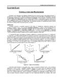
8 It is critical to understand the different activators that may be used for THROMBOELASTOGRAPHY to induce the clotting cascade. Conventional TEG is activated with kaolin, which initiates the intrinsic pathway, resulting in the reaction time or R-time that is similar to a PTT. Rapid THROMBOELASTOGRAPHY utilizes tissue factor (TF) in addition to kaolin, activating both the intrinsic and extrinsic pathways, producing the activated clotting time or TEG-ACT. As the clot begins to form in the presence of the activator, the TEG records the visceroelastic changes that occur in the sample.
9 A pin suspended in the blood within a stationary cup is monitored for motion as fibrin-platelet bonding occurs. This motion is related to the strength of the clot and is analyzed by a computer, which creates a graphical representation. As noted, coagulation factors begin to accumulate producing the R-time or TEG-ACT. These values will be prolonged in the presence of coagulopathy and shortened in hypercoagulability. At this point, fibrin cross-linking begins the initial phases of clot formation. This is measured as both a time index known as the K-time (defined as time to 20 mm of clot) and as a rate.
10 This rate can be determined by the slope of the TEG tracing and is expressed as the -angle. The K-time and -angle thus reflect the levels of circulating fibrinogen. Platelet aggregation then occurs until maximum clot strength occurs at the maximum amplitude (MA). Lysis of the clot may then begin, especially in the presence of severe coagulopathy or of fibrinolytic agents. The percentage of clot that has dissipated at 30 minutes time is expressed as the LY-30. The figure to the left shows the correlation between the coagulation cascade and TEG. tracing, and are summarized at the end of this document.