Scanning Electron Microscope
electron source, optical aberrations of the final lens (objective) and the diffraction aberration on the final aperture. The spot size is smaller at shorter working distances. The incident electron beam is cone-shaped. The vertex angle of the cone is determined by the aperture angle. The wider the cone, the lower the depth of focus.
Download Scanning Electron Microscope

Information
Domain:
Source:
Link to this page:
Documents from same domain

Classification - ccmr.cornell.edu
www.ccmr.cornell.eduwhole organizational system for living things is called “classification.” It works like this: Suppose you wanted to classify a car. See Figure 3. The first group, or kingdom, would all be cars. That would exclude trucks, school buses, and RVs. The kingdom of cars would be further divided into all sedans.

Buoyancy - Cornell Center for Materials Research
www.ccmr.cornell.eduThe Archimedes Principle The mathematician Archimedes, who lived in the third century B.C., is credited with discovering much of how buoyancy works.

Non-Destructive Testing Reading
www.ccmr.cornell.eduLeak Testing (LT) Several techniques are used to detect and locate leaks in pressure containment parts, pressure vessels, and structures. Leaks can be detected by using electronic listening devices, pressure gauge measurements, liquid and gas penetrant techniques, or simple soapbubble tests.

Trace Evidence – How Fibers and Hair are used to aid in ...
www.ccmr.cornell.eduTrace Evidence – How Fibers and Hair are used to aid in Crime Solving. Locard’s Exchange Principal – when a criminal comes in contact with a person or object a cross ... Medullas have three general appearances, as well as many different shapes. Deer Medulla (notice it is as thick as the hair) Rabbit Medulla (again the medulla is very large)

WHAT IS THE SCIENTIFIC METHOD?
www.ccmr.cornell.eduterms of combinations of air, earth, fire and water. One French scientist, Antoine Lavoisier, (1743-1794) worked to change the way chemistry was done. Lavoisier had a habit of making very careful measurements in his experiments. One of the things he discovered from these experiments was that combustion is the result of a reaction with oxygen.
DNA: Definition, Structure, and Discovery
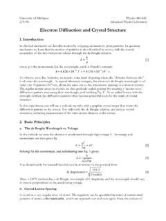
www.ccmr.cornell.edustructure of nucleic acids and i t s signi f i cance f or i nf ormat i on t ransf er i n living material." DNA sequencing DNA sequencing is t echnol ogy t hat al l ows researchers t o det ermi ne t he order of bases in a D NA sequence. T he t echnol ogy can be used t o determine the order of bases i n genes, chromosomes, or an ent i re ...
Related documents
Electron Diffraction and Crystal Structure
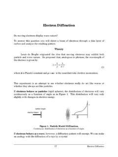
instructor.physics.lsa.umich.eduthe funny story). Thompson, who verified that the electron was a wave, was the son of J.J. Thompson, who discovered that the electron was a particle! Davison and Thompson got the Nobel Prize in 1937. d. The “Powder Method” The Bragg picture tells us that a beam of fixed wavelength (i.e. fixed energy) striking a crystal at the
Electron Diffraction - Boston University
physics.bu.eduelectron beam. The beam will have kinetic energy equal to the change in electric potential energy (eVa). If the beam velocity is non–relativistic (eVa<<mc2), we have at the anode: 1 2 mv 2 = eVa (3) CARBON TARGET As the electron beam passes into the anode, it strikes a very fine nickel screen which holds vaporized graphite (carbon).

Introduction to the Scanning Electron Microscope
imf.ucmerced.eduan electron beam because electrons will quickly disperse or scatter due to collisions with other molecules. 2. Electron beam generation system. This system is found at the top of the microscope column (Fig. 1). This system generates the "illuminating" beam of electrons known as the primary (1 o) electron beam. 3. Electron beam manipulation system.

Measurement of Charge-to-Mass (e/m) Ratio for the Electron
ixnovi.people.wm.eduThe electron beam leaves a visible trail in the tube, because some of the electrons collide with helium atoms, which are excited and then radiate visible light. The electron gun is shown in Fig. 2b. The heater heats the cathode, which emits electrons. …
Optical Microscope; • Scanning Electron Microscope (SEM ...
my.eng.utah.edu• Electron wave is a unique medium that can be used in imaging. By accelerating the electrons into high energy beam (via high voltage), the wavelength thus created is far shorter than white light. For example, for an electron beam produced from a 20 kV gun, the wavelength is only 1240.7/20,000 (eV) = 0.06 nm = 0.6 Å,

Microscopy I Light and Electron Microscopy
www.auburn.eduThe electron beam is absorbed or deflected by the heavy metal stains and shadows are cast onto film or a phosphorescent plate (image is a shadow) at the bottom of the column. - 2-D image - reveals internal cell structure - high resolution, high magnification
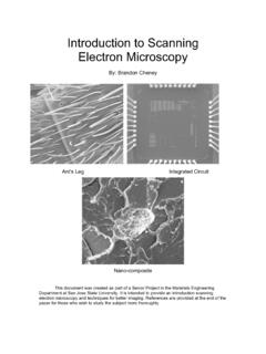
Introduction to Scanning Electron Microscopy
www.sjsu.eduFeb 01, 2005 · The electron beam should have a circular cross section when it strikes the specimen however it is usually elliptical thus the stigmator acts to control this problem [1,4]. 5) Specimen Chamber: At the lower portion of the column the specimen stage and controls are located. The secondary electrons from the
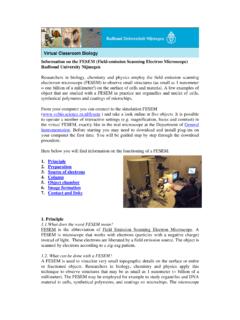
Information on the FESEM (Field-emission Scanning Electron ...
www.vcbio.science.ru.nlThe electron beam is focused by the electro-magnetic lenses (condenser lens, scan coils, stigmator coils and objective lens) and the apertures in the column to a tiny sharp spot. 1. Condenser lens The current in the condenser determines the diameter of the beam: a low current results in a small diameter, a higher current in a larger beam. A narrow