Scanning Electron Microscope
IntroductionMIRA 3 FEG 3 -SEM 1 Introduction The MIRA 3 series is a family of high-quality; fully PC-controlled scanning electron microscopes equipped with a Schottky Field Emission electron gun designed for high vacuum
Download Scanning Electron Microscope

Information
Domain:
Source:
Link to this page:
Documents from same domain

Classification - ccmr.cornell.edu
www.ccmr.cornell.eduwhole organizational system for living things is called “classification.” It works like this: Suppose you wanted to classify a car. See Figure 3. The first group, or kingdom, would all be cars. That would exclude trucks, school buses, and RVs. The kingdom of cars would be further divided into all sedans.

Buoyancy - Cornell Center for Materials Research
www.ccmr.cornell.eduThe Archimedes Principle The mathematician Archimedes, who lived in the third century B.C., is credited with discovering much of how buoyancy works.

Non-Destructive Testing Reading
www.ccmr.cornell.eduLeak Testing (LT) Several techniques are used to detect and locate leaks in pressure containment parts, pressure vessels, and structures. Leaks can be detected by using electronic listening devices, pressure gauge measurements, liquid and gas penetrant techniques, or simple soapbubble tests.

Trace Evidence – How Fibers and Hair are used to aid in ...
www.ccmr.cornell.eduTrace Evidence – How Fibers and Hair are used to aid in Crime Solving. Locard’s Exchange Principal – when a criminal comes in contact with a person or object a cross ... Medullas have three general appearances, as well as many different shapes. Deer Medulla (notice it is as thick as the hair) Rabbit Medulla (again the medulla is very large)

WHAT IS THE SCIENTIFIC METHOD?
www.ccmr.cornell.eduterms of combinations of air, earth, fire and water. One French scientist, Antoine Lavoisier, (1743-1794) worked to change the way chemistry was done. Lavoisier had a habit of making very careful measurements in his experiments. One of the things he discovered from these experiments was that combustion is the result of a reaction with oxygen.
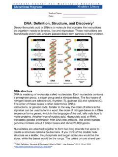
DNA: Definition, Structure, and Discovery
www.ccmr.cornell.edustructure of nucleic acids and i t s signi f i cance f or i nf ormat i on t ransf er i n living material." DNA sequencing DNA sequencing is t echnol ogy t hat al l ows researchers t o det ermi ne t he order of bases in a D NA sequence. T he t echnol ogy can be used t o determine the order of bases i n genes, chromosomes, or an ent i re ...
Related documents

AN INTRODUCTION TO THE COMPOUND MICROSCOPE
users.rowan.eduINTRODUCTION: The light microscope can extend our ability to see detail by 1000 times, so that we can see objects as small as 0.1 micrometer (um) or 100 nanometers (nm) in diameter. The transmission electron microscope extends this capability to objects as small as 0.5 nm in diameter, 1/200,000th the size of objects that are visible to the ...

Introduction to Ethical Studies
philosophy.lander.eduIntroduction to Ethical Studies An Open Source Reader Lee Archie John G. Archie

Introduction to Liquid Crystals
uh.eduIntroduction to Liquid Crystals The study of liquid crystals began in 1888 when an Austrian botanist named Friedrich Reinitzer observed that a material known as cholesteryl benzoate had two distinct melting points. In his experiments, Reinitzer increased the temperature of a solid sample and watched the crystal change into a hazy liquid.

Introduction to the Scanning Electron Microscope
imf.ucmerced.eduscanning electron microscope (SEM). The course is designed as an introduction to the SEM and as a research tool for students who have had no previous SEM experience. Objectives of the course are to define and illustrate the major components of the SEM, as well as describe methodology of operation.
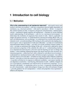
1 Introduction to cell biology - Stanford University
biomechanics.stanford.edu1 Introduction to cell biology 1.1 Motivation ... hoek was the first person to ever observe a cell under a microscope in 1674. The cell theory biologists use nowadays dates back to major contributions of Schwann and Schleiden in 1839, enhanced by contributios of Virchow in 1858.
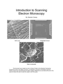
Introduction to Scanning Electron Microscopy
www.sjsu.eduFeb 01, 2005 · Introduction to Scanning Electron Microscopy By: Brandon Cheney Ant’s Leg Integrated Circuit Nano-composite This document was created as part of a Senior Project in the Materials Engineering Department at San Jose State University. It is intended to provide an introduction scanning

All you wanted to know about Electron Microscopy
web.pdx.eduIntroduction This booklet is written for those who know little or nothing about electron microscopy and would like to know how an electron microscope works, why it is used and what useful results it can produce. "With a microscope you see the surface of things. It magnifies them but does not show you reality. It makes things seem higher and wider.
Introduction to microstructure - Inference
www.inference.org.ukIntroduction to microstructure 1.1 What is microstructure? When describing the structure of a material, we make a clear distinction between its crystal ... Essentially the microscope is a tube whose diameter puts a maximum limit on the orders of diffraction maxima which manage to exit the tube to form an image. Making the tube wider

Biology 1290B: An introduction to general microbiology. 1 ...
instruct.uwo.caBiology 1290B Lecture Notes 1-7 2 Key figures in the history of microbiology Robert Hooke (1635 - 1703) was a “polymath’ he made many scientific discoveries in the 17 th century, including making one of the first microscopes and also using a copy of one of Leeuwenhoek’s microscopes to see and draw details of the structure of plant cells