Transcription of CARDIOLOGY - University of Alberta
1 CARDIOLOGYDr. A. WooStephen Juvet, Daniel Kreichman and Manish Sood, chapter editorsKatherine Zukotynski, associate editorBASIC CLINICAL CARDIOLOGY EXAM ..2 Cardiac HistoryFunctional Classification of Cardiovascular DisabilityCardiac ExaminationCARDIAC DIAGNOSTIC TESTS ..6 ECG Interpretation - The BasicsHypertrophy and Chamber EnlargementIschemia/InfarctionMiscellane ous ECG ChangesAmbulatory ECG (Holter Monitor)Echocardiography (2-D ECHO)Coronary AngiographyCardiac Stress Tests and Nuclear CardiologyTests of Left Ventricular (LV) FunctionARRHYTHMIAS.
2 12 Mechanisms of ArrhythmiasAltered Impulse FormationAltered Impulse ConductionOther Etiologic FactorsClinical Approach to Arrhythmias BradyarrhythmiasConduction DelaysTachyarrhythmiasSupraventricular Tachyarrhythmias (SVT s)Ventricular Tachyarrhythmias (VT s)Pre-excitation SyndromesPacemaker IndicationsPacing TechniquesISCHEMIC HEART DISEASE ..19 BackgroundAngina PectorisAcute Coronary SyndromesUnstable Angina/Non ST Elevation Myocardial Infarction (MI)Acute ST Elevation MISudden DeathHEART FAILURE ..27 Compensatory ResponsesSystolic vs. Diastolic DysfunctionSleep-Disordered BreathingHigh-Output Heart FailureAcute Cardiogenic Pulmonary EdemaCardiac TransplantationMCCQE 2002 Review NotesCardiology C1 CARDIOMYOPATHIES.
3 32 Dilated Cardiomyopathy (DCM)Hypertrophic Cardiomyopathy (HCM)Restrictive Cardiomyopathy (RCM)MyocarditisVALVULAR HEART DISEASE ..36 Infective Endocarditis (IE)Rheumatic FeverAortic Stenosis (AS)Aortic Regurgitation (AR)Mitral Stenosis (MS)Mitral Regurgitation (MR)Mitral Valve ProlapseTricuspid Valve DiseasePulmonary Valve DiseaseProsthetic ValvesPERICARDIAL DISEASE ..45 Acute PericarditisPericardial EffusionCardiac TamponadeConstrictive PericarditisSYNCOPE ..47 EVIDENCE-BASED CARDIOLOGY ..48 Congestive Heart Failure (CHF)Ischemic Heart Disease (IHD)Atrial Fibrillation (A fib)COMMONLY USED CARDIAC.
4 49 MEDICATIONS -blockersCalcium Channel Blockers (CCB)Angiotensin Converting Enzyme (ACE) InhibitorsAngiotensin II BlockersDiureticsNitratesAnti-Arrhythmic Anti-PlateletREFERENCES ..52C2 CARDIOLOGY MCCQE 2002 Review NotesBASIC CLINICAL CARDIAC EXAMCARDIAC HISTORY coronary artery disease: chest pain (CP) (location, radiation, duration, intensity, activities associated with onset; alleviating factors (associated with rest, NTG) heart failure: fatigue, presyncope left-sided symptoms: decreased exercise tolerance, shortness of breath on extertion (SOBOE)/chest pain on exertion (CPOE) right-sided symptoms: paroxysmal nocturnal dyspnea (PND)/orthopnea, SOB at rest, ascites, Arrhythmia: presyncopal/syncopal episodes, palpitations Baseline function.)
5 Exercise tolerance (# flights of stairs/blocks), need for nitroglycerin (NTG), symptoms during low impact activities/daily activities (combing hair, showering) or at restFUNCTIONAL CLASSIFICATION OF CARDIOVASCULAR DISABILITYT able 1. Canadian Cardiovascular Society (CCS) Functional ClassificationClassFunctionIordinary physical activity does not cause angina; angina only with strenuous or prolonged activityIIslight limitation of physical activity; angina brought on at > 2 blocks on level (and/or by emotional stress)IIImarked limitation of physical activity; angina brought on at 2 blocks on levelIVinability to carry out any physical activity without discomfort; angina may be present at restTable 2.
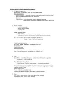
6 New York Heart Association (NYHA) Functional ClassificationClassFunctionIordinary physical activity does not evoke symptoms (fatigue, palpitation, dyspnea, or angina)IIslight limitation of physical activity; comfortable at rest; ordinary physical activity results in symptomsIIImarked limitation of physical activity; less than ordinary physical activity results in symptomsIVinability to carry out any physical activity without discomfort; symptoms may be present at restCARDIAC EXAMINATIONG eneral Examination Skin peripheral vs. central cyanosis, clubbing, splinter hemorrhages, Osler s nodes, Janeway lesionsbrownish-coloured skin hemochromatosis Eyes conjunctival hemorrhages, Roth spots, emboli, copperwire lesions, soft/hard exudatesBlood Pressure (BP) should be taken in both arms with the patient supine and upright be wary of calcification of the radial artery in the elderly as it may factitiously elevate BP (Osler s sign)
7 Orthostatic hypotension - postural drop > 20 mm Hg systolic or > 10 mm Hg diastolic increased HR > 30 bpm (most sensitive - implies inadequate circulating volume)patient unable to stand - specific sign for significant volume depletion pulse pressure(PP) (PP = systolic BP (SBP) - diastolic PB (DBP)) wide PP: increased cardiac output (CO) (anxiety, exercise, fever, thyrotoxicosis, AR, HTN),decreased total peripheral resistance (TPR) (anaphylaxis, liver cirrhosis, nephrotic syndrome, AVM) narrow PP: decreased CO (CHF, shock, hypovolemia, acute MI, hypothyroidism, cardiomyopathy),increased TPR (shock, hypovolemia), valvular disease (AS, MS, MR), aortic disease ( coarctation of aorta) pulsus paradoxus (inspiratory drop in SBP > 10 mmHg ).
8 Cardiac tamponade, constrictive pericarditis, airway obstruction, superior vena cava (SVC) obstruction, COPD (asthma, emphysema)The Arterial Pulse remark on rate, rhythm, volume/amplitude, contour amplitude and contour best appreciated in carotid arteries pulsus alternans - beat-to-beat alteration in PP amplitude with cyclic dip in systolic BP; due to alternating LV contractile force (severe LV dysfunction) pulsus parvus et tardus slow uprising of the carotid upstroke due to severe aortic stenosis (AS) pulsus bisferiens a double waveform due to AS + AR combined spike and dome pulse double carotid impulse due to hypertrophic obstructive cardiomyopathy (HOCM)MCCQE 2002 Review NotesCardiology C3 BASIC CLINICAL CARDIAC EXAM.
9 Inspection observe for apex beat, heaves, liftsPrecordial Palpation apex - most lateral impulse PMI - point of maximal intensity location: normal at 5th intraclavicular space (ICS) at midclavicular line ( 10 cm from midline), lateral/inferior displaced in dilated cardiomyopathy (DCS) size : normal is 2-3 cm in diameter, diffuse > 3 cm duration: normal is <1/2 systole (duration > 2/3 systole is considered sustained) amplitude (exaggerated, brief - AR, MR, L to R shunt) morphology (may have double/triple impulse in HOCM) abnormal impulses palpable heart sounds ( S1 in MS, P2 = pulmonary artery (PA) pulsation, S3, S4) left parasternal lift (right ventricular enlargement (RVE), left atrial enlargement (LAE) , severe left ventricular hypertrophy (LVH)) epigastric pulsation (RVH especially in COPD) thrills (tactile equivalents of murmurs)
10 Over each valvular areaClinical Pearl Left parasternal lift - DDx - RVH (with pulmonary hypertension (HTN), LAE (secondary to severe MR), severe LVH, rarely thoracic aortic - Heart Sounds S1 composed of audible mitral (M1) and tricuspid (T1) components may be split in the normal young patient if S1is loud short PR interval high left atrial (LA) pressure ( early MS) high output states or tachycardia (diastole shortened) if S1is soft first degree AV block calcified mitral valve (MV) ( late MS) high LV diastolic pressures ( CHF, severe AR) occasionally in MR if S1varies in volume AV dissociation (complete AV block, ventricular tachycardia (VT)) AFib S2 normally has 2 components on inspiration.)
