Transcription of Cardiovascular System: Heart - Western Oregon University
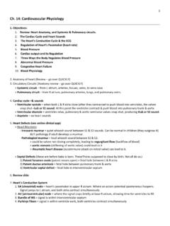
1 1 Cardiovascular System: Heart Roughly size of human fist (~ 250 350 grams) Heart : Double pump composed of cardiac muscle Located in the mediastinum (medial cavity of thorax) Apex Marieb & Hoehn (Human Anatomy and physiology , 8th ed.) Figures / 2/3 of Heart mass lies left of mid-sternal line Base Point of maximum intensity Cardiovascular System Heart Coronary sulcus Anterior intraventricular sulcus 2 Pulmonary trunk Left atrium Pericardial sac: Double-walled sac enclosing the Heart Fibrous pericardium Protects Heart Anchors Heart Prevents overfilling Parietal pericardium Visceral pericardium Contains serous fluid (friction-free environment) Pericardial cavity Pericarditis: Inflammation of the pericardial sac Cardiovascular System Heart Heart : Marieb & Hoehn (Human Anatomy and physiology , 8th ed.)
2 Figure Cardiac Tamponade: Compression of Heart due to fluid / blood build up in pericardial cavity Pulmonary trunk Left atrium Heart layers: Epicardium Myocardium Anchors cardiac fibers Reinforces Heart structures Directs electrical signals Endocardium Cardiovascular System Heart Heart : Often infiltrated with fat Contains fibrous skeleton Marieb & Hoehn (Human Anatomy and physiology , 8th ed.) Figures / 3 Heart Chambers, Vessels & Valves Atria: Receiving chambers Small, thin-walled Right atrium Ventricles: Discharging chambers Large, thick-walled Right ventricle Cardiovascular System Heart Auricles: ( little ears ) Increase atrial volume Pectinate muscles: Muscle bundles; assist in atrial contraction Left atrium Fossa ovalis: Shallow depression; remnants of hole between atria in fetal Heart Left ventricle Trabeculae carneae: Muscle ridges; assist in maintaining momentum Papillary muscle: Cone-like muscle; assists in valve closure Ligamentum arteriosum: Remnant of fetal duct between aorta and pulmonary truck (ductus arteriosus) Ligamentum arteriosum Marieb & Hoehn (Human Anatomy and physiology , 8th ed.)
3 Figure Superior vena cava Pulmonary trunk Aorta Pulmonary veins (4) Returns blood above diaphragm Inferior vena cava Returns blood below diaphragm Carries blood to lungs Returns blood from lungs Carries blood to body (largest artery in body) Heart Chambers, Vessels & Valves Cardiovascular System Heart Marieb & Hoehn (Human Anatomy and physiology , 8th ed.) Figure 4 Side-by-side pumps of the Heart serve separate circuits Pulmonary circuit Systemic circuit In steady state, cardiac output from each ventricle must be equal as well as the venous return to each atrium O2-poor; CO2-rich O2-rich; CO2-poor Heart Chambers, Vessels & Valves Cardiovascular System Heart Right side Left side Workload not equal (short, low pressure loop) (long, high pressure loop) Costanzo ( physiology , 4th ed.)
4 Figure O2-poor; CO2-rich O2-rich; CO2-poor Blood in the atria / ventricles provides little nourishment to Heart tissue How does the Heart itself get nourishment? Right coronary artery Answer: Coronary circulation Left coronary artery Anterior interventricular artery Angina pectoris: Thoracic pain caused by fleeting deficiency in blood delivery Myocardial infarction: Myocardial cell death resulting from prolonged coronary blockage ( Heart attack) Heart Chambers, Vessels & Valves Cardiovascular System Heart Right marginal artery Posterior interventricular artery Circumflex artery (feeds right lateral ventricular wall) (feeds posterior ventricular walls) (feeds left atrium and posterior wall of left ventricle) (feeds interventricular septum and anterior ventricular walls) 1/200 body s mass; 1/20 body s blood supply Anastomosis (junction of vessels) Marieb & Hoehn (Human Anatomy and physiology , 8th ed.)
5 Figure 5 Blood in the atria / ventricles provides little nourishment to Heart tissue How does the Heart itself get nourishment? Coronary sinus Answer: Coronary circulation Heart Chambers, Vessels & Valves Cardiovascular System Heart Great cardiac vein (empties blood back into right atrium) Middle cardiac vein Small cardiac vein Coronary circulation delivery limited to when Heart is Marieb & Hoehn (Human Anatomy and physiology , 8th ed.) Figure Right atrioventricular valve (tricuspid valve) Blood flows through the Heart in a single direction due to the presence of valves Left atrioventricular valve (bicuspid valve) (mitral valve) Pulmonary semilunar valve Aortic semilunar valve Cardiovascular System Heart Heart Chambers, Vessels & Valves Cusp: Flap of endocardium reinforced by connective tissue core Marieb & Hoehn (Human Anatomy and physiology , 8th ed.)
6 Figure 6 Atrioventricular valves (prevent backflow into atria) Papillary muscle Chordae tendineae Semilunar valves (prevent backflow into ventricles) Valves open / close based on pressure differences (collagen cords) (papillary muscles tighten) Marieb & Hoehn (Human Anatomy and physiology , 8th ed.) Figures / Cardiovascular System Heart Heart Chambers, Vessels & Valves Cardiovascular System Heart Heart Chambers, Vessels & Valves Aortic semilunar valve (pig) Pathophysiology: Valvular Regurgitation: Valve does not close properly; blood regurgitated Valvular stenosis: Valve flaps become stiff; opening constricted Treatment = Valve replacement Causes: Congenially deformed valve Post-inflammatory scarring Infective endocarditis Rupture of cord / muscle Causes: Congenially deformed valve Post-inflammatory scarring Calcification of valve 7 Heart designed to create complex flow patterns (direct / maintain blood momentum) Randall et al.
7 (Animal physiology , 5th ed.) Figures / 1) Chambers arranged in loop pattern 2) Delivery vessels curved 3) Grooves / ridges within chambers Cardiovascular System Heart Heart Chambers, Vessels & Valves Cardiovascular System Heart Muscle Fiber Anatomy Marieb & Hoehn (Human Anatomy and physiology , 8th ed.) Electrical synapses (intercalated discs) Functional syncytium: The entire myocardium behaves as a single coordinated unit Striated, branched cells (~ 85 100 m) Single nucleus (sometimes ) Large [mitochondria] (~ 15x skeletal muscle) High fatigue resistance Desmosome Gap junction Less elaborate T-tubule system and sarcoplasmic reticulum compared to skeletal muscle Contractile cell: 8 Conducting cells: Cardiac cells specialized to quickly spread action potentials across myocardium Weak force generators Cardiovascular System Heart Intrinsic Conduction System: Marieb & Hoehn (Human Anatomy and physiology , 8th ed.
8 Figure Sinoatrial node: (SA node) Located in right atrial wall Initiates action potentials (APs) Pacemaker (~ 80 beats / min) Atrial internodal tracts Atrioventricular node: (AV Node) Connects atria to ventricles Slowed conduction velocity Ventricular filling Bundle of His Bundle branches Purkinje fibers Normal sinus rhythm: 1) AP originates at SA node 2) SA node fires at 60 100 beats / min 3) Correct myocardial activation sequence System allows for orderly, sequential depolarization and contraction of Heart Cardiac Electrophysiology Cardiovascular System Heart Marieb & Hoehn (Human Anatomy and physiology , 8th ed.) Figure The concepts applied to cardiac APs are the same concepts as applied to APs in nerves / skeletal muscle Review: Membrane potential determined by relative conductances / concentrations of permeable ions Ions flow down electrochemical gradient toward equilibrium potential (Nernst equation) Membrane potential expressed in mV.
9 Inside cell expressed relative to outside Resting membrane potential determined primarily by K+ ions (leaky K+ gates at rest) Na+ / K+ pumps maintain [gradients] across membranes Changes in membrane potential caused by flow of ions into / out of cell Threshold potential represents the point at which a depolarization even becomes self-sustaining (voltage-gated channels) Cardiac Electrophysiology 9 Cardiovascular System Heart APs of Atria, Ventricles & Purkinje System: Time (ms) Tension (g) Membrane potential (mV) Marieb & Hoehn (Human Anatomy and physiology , 8th ed.) Figure Long duration AP (150 300 ms) Long refractory period Maximum Heart rate: ~ 240 beats / min Stable RMP Plateau: Sustained period of depolarization Costanzo ( physiology , 4th ed.)
10 Figure Cardiac Electrophysiology Cardiovascular System Heart APs of Atria, Ventricles & Purkinje System: Costanzo ( physiology , 4th ed.) Figure Phases of the Action Potential: Phase 0 Upstroke Period of rapid depolarization Na+ enters via VG channels ( gNa) gX = conductance Phase 1 Initial repolarization Phase 2 Plateau Stable, depolarized membrane potential K+ exits via VG channels ( gK) Ca++ enters via VG channels ( gCa) Brief period of repolarization Na+ channels close ( gNa) K+ exits via VG channels ( gK) Steep electrochemical gradient VG = voltage-gated Net current = 0 Ca2+ entry initiates release of more Ca2+ from intracellular stores (excitation-contraction coupling) (L-type) Cardiac Electrophysiology 10 Cardiovascular System Heart APs of Atria, Ventricles & Purkinje System: Costanzo ( physiology , 4th ed.)