Transcription of CHAPTER 8 Identification and Characterization of ...
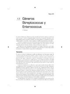
1 1 CHAPTER 8 Identification and Characterization of streptococcus pneumoniae S. pneumoniae may occur intracellularly or extracellularly as gram-positive lanceolate diplococci, but can also occur as single cocci or in short chains of cocci. S. pneumoniae is a fastidious bacterium, growing best at 35-37 C with ~5% CO2 (or in a candle-jar). It is usually cultured on media that contain blood, but can also grow on a chocolate agar plate (CAP). On a blood agar plate (BAP), colonies of S. pneumoniae appear as small, grey, moist (sometimes mucoidal), colonies and characteristically produce a zone of alpha-hemolysis (green) (Figure 1). The alpha-hemolytic property differentiates this organism from many species, but not from the commensal alpha-hemolytic (viridans) streptococci.
2 Differentiating pneumococci from viridans streptococci is difficult as young pneumococcal colonies appear raised, similar to viridans streptococci. However, once the pneumococcal culture ages 24-48 hours, the colonies become fl attened, and the central portion becomes depressed, which does not occur with viridans streptococci (Figure 2). A microscope (30-50X) or a 3X hand lens can also be a useful tool in differentiating pneumococci from viridans streptococci. Prior to Identification and Characterization testing procedures, isolates should always be inspected for purity of growth and a single colony should be re-streaked, when necessary, to obtain a pure culture. For the following Identification and Characterization procedures, i t is essential to test alpha-hemolytic colonies that are less than a day old, typically grown overnight at 35-37 C with ~5% CO2 (or in a candle-jar).
3 The following specialized tests are used to identify colonies on a BAP that resemble pneumococci (Figure 3). S. pneumoniae can be identified using Gram stain, catalase, and optochin tests simultaneously, with bile solubility as a confirmatory test. If these tests indicate that the isolate is S. pneumoniae , serological tests to identify the serotype can be performed. This sequence of testing is an efficient way to save costly serotyping reagents and time. Additional methods for Identification and Characterization of S. pneumoniae using molecular tools are described in CHAPTER 10: PCR Methods and CHAPTER 12: Molecular Methods. Additional protocols used for streptococcal species Identification and updates to existing methods can be found at: Biosafety Level 2 (BSL-2) practices are required for work involving isolates of S.
4 pneumoniae , as this organism presents a potential hazard to laboratory personnel and the surrounding working environment. Please refer to CHAPTER 4: Biosafety in order to follow the guidelines that have been established for laboratorians working in BSL-2 facilities as many of the tests described in this CHAPTER require opening plates with live cultures and are often performed outside of a biosafety cabinet (BSC). 2 Figure 1. S. pneumoniae colonies with a surrounding green zone of alpha-hemolysis (black arrow) on a BAP Figure 2. S. pneumoniae colonies have a flattened and depressed center after 24-48 hours of growth on a BAP, whereas the viridans streptococci retain a raised center 3 Plate on blood agar All 3 tests must be performed in parallel Determine serotype Report as S.
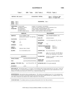
5 pneumoniae Gram stain Specimen (CSF/blood) Optochin test Catalase test Identify alpha-hemolytic colonies Identify as probable streptococcus spp. from Gram stain and catalase result Report as S. pneumoniae (assuming it is Gram-positive, catalase-negative) Bile solubility test Catalase negative Identify gram-positive diplococci or gram-positive cocci in short chains Bile soluble Bile insoluble Optochin resistant (<14 mm diameter) Optochin susceptible ( 14 mm diameter) -hemolytic streptococci other than S. pneumoniae 4 Figure 3. Flow chart for Identification and Characterization of a S. pneumoniae isolate I. Catalase t est Catalase is the enzyme that breaks down hydrogen peroxide (H2O2) into H2O and O2.
6 The oxygen is given off as bubbles in the liquid. The catalase test is primarily used to differentiate between gram-positive cocci. Members of the genus Staphylococcus are catalase-positive, and members of the genera streptococcus and Enterococcus are catalase-negative. A. Performing the catalase test 1. Grow the isolate(s) to be tested for 18-24 hours on a BAP at 35-37 C with ~5% CO2 (or in a candle-jar). 2. From overnight growth on the BAP, use a disposable loop to carefully remove a colony and place it on a glass slide. Do not transfer any of the blood agar to the slide as erythrocytes in the blood agar will cause a false-positive reaction. 3. Add ml of 3% H2O2 to the slide and mix with the bacteria.
7 H2O2 can be obtained from a commercial drug store. After initially opening, store H2O2 at 4 C in a tightly closed bottle as it will slowly lose potency once opened. 4. Observe the bacterial suspension on the slide immediately for vigorous bubbling. 5. It is essential to use a known positive and negative quality control (QC) strain. A Staphylococcus spp. strain can be used for a positive control and a known S. pneumoniae strain or any other streptococcal spp., , S. pyogenes can be used for a negative control. B. Reading the catalase test results The absence of bubbling from a transferred colony indicates a negative test. Any bubbling from a transferred colony indicates a positive test (Figure 4).
8 C. Troubleshooting False positives will result from transfer of red blood cells so take care when picking colonies from the BAP for this test. 5 D. Quality control It is essential to use a known positive and negative QC strain as described in the procedure. Opened bottles should be checked against a known catalase positive organism every 6 months. Figure 4. Negative and positive catalase test results. The absence of bubbling from a transferred colony indicates a negative test. All streptococci are catalase-negative. II. Optochin test S. pneumoniae strains are sensitive to the chemical optochin (ethylhydrocupreine hydrochloride). Optochin sensitivity allows for the presumptive Identification of alpha-hemolytic streptococci as S.
9 pneumoniae , although some pneumococcal strains are optochin-resistant. Other alpha-hemolytic streptococcal species are optochin-resistant. A. Performing the optochin test Optochin (P) disks (6 mm, 5 g) can be obtained from a commercial vendor. Optochin disks are often called P disks and many commercial versions are labeled with a capital P . If a commercial source of P disks is not available, a 1:4000 solution of ethylhydrocupreine hydrochloride can be applied to sterile 6 mm filter paper disks. 1. Grow the strain(s) to be tested for 18-24 hours on a BAP at 35-37 C w ith ~5% CO2 (or in a candle-jar). 2. Use a disposable loop to remove an isolated colony from the overnight culture on the BAP and streak onto one half of a BAP.
10 6 Two different isolates can be tested on the same plate, but care must be taken to ensure that the cultures do not overlap. 3. Place a P disk within the streaked area of the plate and incubate the BAP overnight at 35-37 C with ~5% CO2 (or in a candle-jar). 4. Observe the growth on the BAP near the P disk and measure the zone of inhibition, if applicable. B. Reading the optochin test results Using a 6 mm, 5 g disk, a zone of inhibition of 14 mm or greater indicates sensitivity and allows for presumptive Identification of pneumococci (Figure 5) . Zones of inhibition should be measured from the top surface of the plate with the top removed. Use either calipers or a ruler with a handle attached for these measurements.