Transcription of Functional Anatomy of the Heart Cardiovascular Physiology
1 1 Cardiovascular PhysiologyLecture Outline Cardiovascular System Function Functional Anatomy of the Heart Myocardial Physiology Cardiac Cycle Cardiac Output Controls & Blood PressureCardiovascular System Function Functional components of the Cardiovascular system: Heart Blood Vessels Blood General functions these provide Transportation Everything transported by the blood Regulation Of the Cardiovascular system Intrinsic v extrinsic Protection Against blood loss Production/SynthesisFunctional Anatomy of the Heart To create the pump we have to examine Cardiac muscle Chambers Valves Intrinsic Conduction System2 Lecture Outline Cardiovascular System Function Functional Anatomy of the Heart Myocardial Physiology Cardiac Cycle Cardiac Output Controls & Blood PressureFunctional Anatomy of the HeartCardiac Muscle Characteristics Striated Short branched cells Uninucleate Intercalated discs T-tubules larger andover z-discsFunctional Anatomy of the HeartChambers 4 chambers 2 Atria 2 Ventricles 2 systems Pulmonary SystemicFunctional Anatomy of the HeartValves Function is to prevent
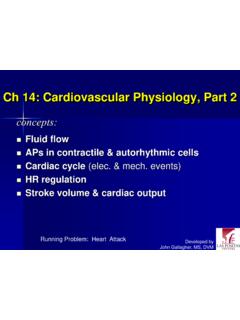
2 Backflow Atrioventricular Valves Prevent backflow to the atria Prolapse is prevented by the chordae tendinae Tensioned by the papillary muscles Semilunar Valves Prevent backflow into ventricles3 Functional Anatomy of the HeartIntrinsic Conduction System Consists of pacemaker cells and conduction pathways Coordinate the contraction of the atria and ventriclesLecture Outline Cardiovascular System Function Functional Anatomy of the Heart Myocardial Physiology Autorhythmic Cells (Pacemaker cells) Contractile cells Cardiac Cycle Cardiac Output Controls & Blood PressureMyocardial PhysiologyAutorhythmic Cells (Pacemaker Cells) Characteristics of Pacemaker Cells Smaller than contractile cells Don t contain many myofibrils No organized sarcomere structure do not contribute to the contractile force of the heartnormal contractile myocardial cellconduction myofibersSA node cellAV node cellsMyocardial PhysiologyAutorhythmic Cells (Pacemaker Cells) Characteristics of Pacemaker Cells Unstable membrane potential bottoms out at -60mV drifts upward to -40mV, forming a pacemaker potential Myogenic The upward drift allows the membrane to reach threshold potential (-40mV) by itself This is due to1.
3 Slow leakage of K+out & faster leakage Na+in Causes slow depolarization Occurs through Ifchannels (f=funny) that open at negative membrane potentials and start closing as membrane approaches threshold potential2. Ca2+channels opening as membrane approaches threshold At threshold additional Ca2+ion channels open causing more rapid depolarization These deactivate shortly after and3. Slow K+channels open as membrane depolarizes causing an efflux of K+and a repolarization of membrane4 Myocardial PhysiologyAutorhythmic Cells (Pacemaker Cells) Characteristics of Pacemaker CellsMyocardial PhysiologyAutorhythmic Cells (Pacemaker Cells) Altering Activity of Pacemaker Cells Sympathetic activity NE and E increase Ifchannel activity Binds to 1adrenergic receptors which activate cAMP and increase Ifchannel open time Causes more rapid pacemaker potential and faster rate of action potentialsSympathetic Activity Summary.
4 Increased chronotropic effects Heart rateincreased dromotropic effects conduction of APsincreased inotropic effects contractilityMyocardial PhysiologyAutorhythmic Cells (Pacemaker Cells) Altering Activity of Pacemaker Cells Parasympathetic activity ACh binds to muscarinic receptors Increases K+permeability and decreases Ca2+permeability = hyperpolarizing the membrane Longer time to threshold = slower rate of action potentialsParasympathetic Activity Summary:decreased chronotropic effects Heart ratedecreased dromotropic effects conduction of APsdecreased inotropic effects contractilityMyocardial PhysiologyContractile Cells Special aspects Intercalated discs Highly convoluted and interdigitatedjunctions Joint adjacent cells with Desmosomes & fascia adherens Allow for synticial activity With gap junctions More mitochondria than skeletal muscle Less sarcoplasmic reticulum Ca2+also influxes from ECF reducing storage need Larger t-tubules Internally branching Myocardial contractions are graded!
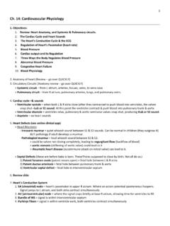
5 5 Myocardial PhysiologyContractile Cells Special aspects The action potential of a contractile cell Ca2+plays a major role again Action potential is longer in duration than a normal action potential due to Ca2+entry Phases4 resting membrane potential @ -90mV0 depolarization Due to gap junctions or conduction fiber action Voltage gated Na+channels close at 20mV1 temporary repolarization Open K+channels allow some K+to leave the cell2 plateau phase Voltage gated Ca2+channels are fully open (started during initial depolarization)3 repolarization Ca2+ channels close and K+ permeability increases as slower activated K+ channels open, causing a quick repolarization What is the significance of the plateau phase?Myocardial PhysiologyContractile Cells Skeletal Action Potential vs Contractile Myocardial Action PotentialMyocardial PhysiologyContractile Cells Plateau phase prevents summation due to the elongated refractory period No summation capacity = no tetanus Which would be fatalSummary of Action PotentialsSkeletal Muscle vs Cardiac Muscle6 Myocardial PhysiologyContractile Cells Initiation Action potential via pacemaker cells to conduction fibers Excitation-Contraction Coupling1.
6 Starts with CICR (Ca2+induced Ca2+release) AP spreads along sarcolemma T-tubules contain voltage gated L-type Ca2+channels which open upon depolarization Ca2+entrance into myocardial cell and opens RyR (ryanodine receptors) Ca2+release channels Release of Ca2+from SR causes a Ca2+ spark Multiple sparks form a Ca2+signalSpark GifMyocardial PhysiologyContractile Cells Excitation-Contraction Coupling2. Ca2+signal (Ca2+from SR and ECF) binds to troponin to initiate myosin head attachment to actin Contraction Same as skeletal muscle, Strength of contraction varies Sarcomeres are not all or none as it is in skeletal muscle The response is graded! Low levels of cytosolic Ca2+will not activate as many myosin/actin interactions and the opposite is true Length tension relationships exist Strongest contraction generated when stretched between 80 & 100% of maximum (physiological range) What causes stretching?
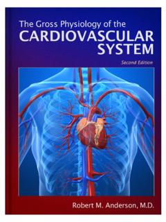
7 The filling of chambers with bloodMyocardial PhysiologyContractile Cells Relaxation Ca2+is transported back into the SR and Ca2+is transported out of the cell by a facilitated Na+/Ca2+exchanger (NCX) As ICF Ca2+levels drop, interactions between myosin/actin are stopped Sarcomere lengthensLecture Outline Cardiovascular System Function Functional Anatomy of the Heart Myocardial Physiology Autorhythmic Cells (Pacemaker cells) Contractile cells Cardiac Cycle Cardiac Output Controls & Blood Pressure7 Cardiac CycleCoordinating the activity Cardiac cycle is the sequence of events as blood enters the atria, leaves the ventricles and then starts over Synchronizing this is the Intrinsic Electrical Conduction System Influencing the rate (chronotropy & dromotropy) is done by the sympathetic and parasympathetic divisions of the ANSC ardiac CycleCoordinating the activity Electrical Conduction Pathway Initiated by the Sino-Atrial node (SA node) which is myogenic at 70-80 action potentials/minute Depolarization is spread through the atria via gap junctions andinternodal pathways to the Atrio-Ventricular node (AV node) The fibrous connective tissue matrix of the Heart prevents further spread of APs to the ventricles A slight delay at the AV node occurs Due to slower formation of action potentials Allows further emptying of the atria Action potentials travel down the Atrioventricular bundle (Bundle of His) which splits into left and right atrioventricular bundles (bundle branches) and then into the conduction myofibers (Purkinje cells)
8 Purkinje cells are larger in diameter & conduct impulse very rapidly Causes the cells at the apex to contract nearly simultaneously Good for ventricular ejectionCardiac CycleCoordinating the activity Electrical Conduction PathwayCardiac CycleCoordinating the activity The electrical system gives rise to electrical changes (depolarization/repolarization) that is transmitted through isotonic body fluids and is recordable The ECG! A recording of electrical activity Can be mapped to the cardiac cycle8 Cardiac CyclePhases Systole = period of contraction Diastole = period of relaxation Cardiac Cycle is alternating periods of systole and diastole Phases of the cardiac cycle1. Rest Both atria and ventricles in diastole Blood is filling both atria and ventricles due to low pressure conditions2. Atrial Systole Completes ventricular filling3. Isovolumetric Ventricular Contraction Increased pressure in the ventricles causes the AV valves to why?
9 Creates the first Heart sound (lub) Atria go back to diastole No blood flow as semilunar valves are closed as wellCardiac CyclePhases Phases of the cardiac cycle4. Ventricular Ejection Intraventricular pressure overcomes aortic pressure Semilunar valves open Blood is ejected5. Isovolumetric Ventricular Relaxation Intraventricular pressure drops below aortic pressure Semilunar valves close = second Heart sound (dup) Pressure still hasn t dropped enough to open AV valves so volume remains same (isovolumetric)Back to Atrial & Ventricular DiastoleCardiac CyclePhases Phases of the cardiac cycle4. Ventricular Ejection Intraventricular pressure overcomes aortic pressure Semilunar valves open Blood is ejected5. Isovolumetric Ventricular Relaxation Intraventricular pressure drops below aortic pressure Semilunar valves close = second Heart sound (dup) Pressure still hasn t dropped enough to open AV valves so volume remains same (isovolumetric)Back to Atrial & Ventricular Diastole9 Cardiac CyclePhasesCardiac CycleBlood Volumes & PressureCardiac CyclePutting it all together!
10 Lecture Outline Cardiovascular System Function Functional Anatomy of the Heart Myocardial Physiology Cardiac Cycle Cardiac Output Controls & Blood next time!