Transcription of g B = -1/2 B - Georgetown University
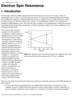
1 electron Spin resonance (ESR) Spectroscopy ( electron paramagnetic resonance , EPR). The principles of ESR are quite analogous to those of NMR. Thus the electron has an intrinsic magnetic moment e = -g eS. where g = , e = eh/4%mc = 10-24J T-1 (Bohr magneton), and S = . The first order Zeeman Effect splits the two ms states of the unpaired electrons in a ms=+1/2. paramagnetic material. g B. ms= -1/2. B. The resonance condition is therefore h = g B, but since the magnetic moment of an electron is 2-3 orders of magnitude greater than that of a magnetic nucleus an ESR spectrometer operates with smaller magnetic fields and higher frequencies than an NMR spectrometer.. Compare a 300 MHz NMR spectrometer with a standard ( X- band ) ESR spectrometer NMR 3 108 Hz and B = Tesla ESR 9 109 Hz and B = Tesla Higher frequency (microwave) radiation requires different technology for ESR spectrometers.
2 The klystron is the source of microwave radiation of fixed . frequency . The cavity (corresponds to the NMR probe) is a hollow rectangular or cylindrical box, the dimensions of which are matched to the wavelength of the microwaves so that the sample (which is ins erted in a quartz nmr-like tube) is held in a region where the magnetic field component of the radiation is maximized and the electric field is minimized. The spectrometer is tuned so that the waveguides and cavity contain standing waves, and the detector records a constant intensity. When the magnetic field is swept to achieve the resonance condition, radiation is absorbed by the sample, and a small decrease in radiation intensity should be observed at the detector. Since it is much more efficient to detect an AC signal in the presence of a large DC background, the magnetic field is modulated (typically at 100 KHz) by means of coils embedded in the walls of the cavity.
3 This generates a signal that looks like the first derivative of the absorption line.. Any sample that contains unpaired electrons can, in principle, yield an ESR (EPR) spectrum. Free radicals (organic and inorganic). triplet states of molecules with diamagnetic ground states transition metal, lanthanide, and actinide complexes with unpaired d or f electrons. Spectra are commonly recorded on solutions or on solids (single crystals, polycrystalline powders, frozed glasses, etc.). The observable characteristics of an ESR spectrum are The position of the signal (analog of the NMR chemical shift). The pattern of electron -nucleus hyperfine coupling (analog of NMR spin coupling). The line-shapes and -widths (relaxation rates, exchange effects, anisotropy as in NMR).. The position of an ESR signal is defined by the effective g- value, by h.
4 G=. eB. g-values are determined by careful measurement of the frequency and magnetic field, or more commonly by using a reference of known g-value. A very common reference is the stable free radical diphenylpicrylhydrazyl (C6H5)2NN(&){C6H2(NO2)3}. for which g = In liquid samples the average (isotropic) value of g is found; in solid samples g is often isotropic (gx , gy , gz). The g-value of a free electron is , and the g-values of most free radicals are very close to this value, since the unpaired electron has very little orbital contribution to the magnetic moment. (carbon-based radicals, spin-orbit coupling very small). So, the g-value of a radical has little significance. On the other hand, g-values for ESR spectra of dn and fn complexes can differ greatly from , because of spin-orbit coupling.
5 Recall that g= is the (anomalous) proportionality between the spin angular momentum and magnetic moment of an electron (in Bohr Magnetons). The corresponding proportionality between the orbital angular momentum and magnetic moment is Thus the effective g for an electron in a dn complex say will depend upon how much orbital angular momentum contributes to the magnetic moment, and whether this orbital contribution acts in parallel or is opposed to the spin angular momentum. Hund's 3rd rule states that the ground state of a dn complex is the one with minimum J (=L-S) if the shell is less than half-full, and maximum J (=L+S) if the shell is more than half full. So for a V4+ (d1) compound we expect g < (typically ). For Cu2+ (d9) g > (typically ). For Mn2+ (d5, L=0) g =.


