Transcription of Human Physiology/The cardiovascular system
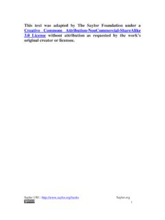
1 Human Physiology/The cardiovascular system1 Human Physiology/The cardiovascular system Blood physiology Human physiology The Immune system Homeostasis Cells Integumentary Nervous Senses Muscular Blood cardiovascular Immune Urinary Respiratory Gastrointestinal Nutrition Endocrine Reproduction (male) Reproduction (female) Pregnancy Genetics Development AnswersModel of a Human heartIntroductionThe heart is the life-giving, ever-beatingmuscle in your chest. From inside thewomb until death, the thump goes heart for the average Human willcontract about 3 billion times; neverresting, never stopping to take a breakexcept for a fraction of a second betweenbeats. At 80 years of age, a person's heartwill continue to beat an average of100,000 times a day. Many believe thatthe heart is the first organ to becomefunctional. Within weeks of conceptionthe heart starts its mission of supplyingthe body with nutrients even though theembryo is no bigger than a capital letteron this page.
2 The primary function of theheart is to pump blood through thearteries, capillaries, and veins. There arean estimated 60,000 miles of vesselsthroughout an adult body. Bloodtransports oxygen, nutrients, diseasecausing viruses, bacteria, hormones andhas other important functions as well. Theheart is the pump that keeps blood circulating properly. Americans today have many options to take care of theirheart and circulatory system . Expanding medical technology has made it much easier to do so. This chapter isdedicated to the heart and its many HeartThe heart is a hollow, muscular organ about the size of a fist. It is responsible for pumping blood through the bloodvessels by repeated, rhythmic contractions. The heart is composed of cardiac muscle, an involuntary muscle tissuethat is found only within this organ. The term "cardiac" (as in cardiology) means "related to the heart and comesfrom the Greek word kardia, for "heart.
3 " It has a four-chambered, double pump and is located in the thoracic cavitybetween the lungs. The cardiac muscle is self-exciting, meaning it has its own conduction system . This is in contrastwith skeletal muscle, which requires either conscious or reflex nervous stimuli. The heart's rhythmic contractionsoccur spontaneously, although the frequency or heart rate can be changed by nervous or hormonal influence such asexercise or the perception of Physiology/The cardiovascular system2 MyocardiumThe myocardium is the muscular tissue of the heart. The myocardium is composed of specialized cardiac musclecells with an ability not possessed by muscle tissue elsewhere in the body. Cardiac muscle, like other muscles, cancontract, but it can also conduct electricity, like nerves. The blood to the myocardium is supplied by the coronaryarteries. If these arteries are occluded by atherosclerosis and/or thrombosis, this can lead to angina pectoris ormyocardial infarction due to ischemia (lack of oxygen).
4 Failure of the heart to contract properly (for various reasons)is termed heart failure, generally leading to fluid retention, edema, pulmonary edema, renal insufficiency,hepatomegaly, a shortened life expectancy and decreased quality of lifePericardiumThe pericardium is the thick, membranous sac that surrounds the heart. It protects and lubricates the heart. There aretwo layers to the pericardium: the fibrous pericardium and the serous pericardium. The serous pericardium is dividedinto two layers; in between these two layers there is a space called the pericardial layer next to the heart is the visceral layer, also known as the Epicardium. This is the innermost layer andconsists of connective ChambersThe heart has four chambers, two atria and two ventricles. The atria are smaller with thin walls, while the ventriclesare larger and much are two atria on either side of the heart.
5 On the right side is the atrium that contains blood which is poor inoxygen. The left atrium contains blood which has been oxygenated and is ready to be sent to the body. The rightatrium receives de-oxygenated blood from the superior vena cava and inferior vena cava. The left atrium receivesoxygenated blood from the left and right pulmonary ventricle is a heart chamber which collects blood from an atrium and pumps it out of the heart. There are twoventricles: the right ventricle pumps blood into the pulmonary circulation for the lungs, and the left ventricle pumpsblood into the systemic circulation for the rest of the body. Ventricles have thicker walls than the atria, and thus cancreate the higher blood pressure. Comparing the left and right ventricle, the left ventricle has thicker walls because itneeds to pump blood to the whole body. This leads to the common misconception that the heart lies on the left sideof the interventricular septum (ventricular septum, or during development septum inferius) is the thick wall separatingthe lower chambers (the ventricles) of the heart from one another.
6 The ventricular septum is directed backward andto the right, and is curved toward the right ventricle. The greater portion of it is thick and muscular and constitutesthe muscular ventricular septum. Its upper and posterior part, which separates the aortic vestibule from the lower partof the right atrium and upper part of the right ventricle, is thin and fibrous, and is termed the membranous Physiology/The cardiovascular system3 ValvesThe two atrioventricular (AV) valves are one-way valves that ensure that blood flows from the atria to the ventricles,and not the other way. The two semilunar (SL) valves are present in the arteries leaving the heart; they prevent bloodfrom flowing back into the ventricles. The sound heard in a heart beat is the heart valves shutting. The right AVvalve is also called the tricuspid valve because it has three flaps. It is located between the right atrium and the rightventricle.
7 The tricuspid valve allows blood to flow from the right atrium into the right ventricle when the heart isrelaxed during diastole. When the heart begins to contract, the heart enters a phase called systole, and the atriumpushes blood into the ventricle. Then, the ventricle begins to contract and blood pressure inside the heart rises. Whenthe ventricular pressure exceeds the pressure in the atrium, the tricuspid valve snaps shut. The left AV valve is alsocalled the bicuspid valve because it has two flaps. It is also known as the mitral valve due to the resemblance to abishop's mitre (liturgical headdress). This valve prevents blood in the left ventricle from flowing into the left it is on the left side of the heart, it must withstand a great deal of strain and pressure; this is why it is made ofonly two cusps, as a simpler mechanism entails a reduced risk of malfunction.
8 There are two remaining valves calledthe Semilunar Valves. They have flaps that resemble half moons. The pulmonary semilunar valve lies between theright ventricle and the pulmonary trunk. The aortic semilunar valve is located between the ventricle and the ApparatusThe chordae tendinae are attached to papillary muscles that cause tension to better hold the valve. Together, thepapillary muscles and the chordae tendinae are known as the subvalvular apparatus. The function of the subvalvularapparatus is to keep the valves from prolapsing into the atria when they close. The subvalvular apparatus have noeffect on the opening and closing of the valves. This is caused entirely by the pressure gradient across the with the HeartThe most common congenital abnormality of the heart is the bicuspid aortic valve. In this condition, instead of threecusps, the aortic valve has two cusps.
9 This condition is often undiagnosed until the person develops calcific aorticstenosis. Aortic stenosis occurs in this condition usually in patients in their 40s or 50s, an average of 10 years earlierthan in people with normal aortic valves. Another common complication of rheumatic fever is thickening andstenosis (partial blocking) of the mitral valve. For patients who have had rheumatic fever dentists are advised toprophylactally administer antibiotics prior to dental work to prevent bacterial endocarditis that occurs when bacteriafrom the teeth enter the circulation and attach to damaged heart aortic valve is a semilunar valve, but it s called bicuspid because of it s regular three "cusps" or "semilunar"valves, and is not to be confused with the left atrioventricular valve, which is more commonly called the mitralvalve, and is one of the two cuspidal Physiology/The cardiovascular system4 Passage of Blood Through the HeartDiagram of the Human heartWhile it is convenient to describe the flowof the blood through the right side of theheart and then through the left side.
10 It isimportant to realize that both atria contractat the same time and that both ventriclescontract at the same time. The heart worksas two pumps, one on the right and one onthe left that works simultaneously. The rightpump pumps the blood to the lungs or thepulmonary circulation at the same time thatthe left pump pumps blood to the rest of thebody or the systemic circulation. Venousblood from systemic circulation(deoxygenated) enters the right atriumthrough the superior and inferior vena right atrium contracts and forces theblood through the tricuspid valve (rightatrioventricular valve) and into the rightventricles. The right ventricles contract andforce the blood through the pulmonarysemilunar valve into the pulmonary trunkand out the pulmonary artery. This takes the blood to the lungs where the blood releases carbon dioxide and receivesa new supply of oxygen.