Transcription of PHYSIOLOGY OF CARDIOVASCULAR SYSTEM - uniba.sk
1 PHYSIOLOGY OF CARDIOVASCULAR SYSTEM PHYSIOLOGY of the Heart Heart rate = 70/min, 100 000/day, 5 1/min, 4 500 l/day Morphology of the heart: 2 separate pumps right/left Each from 2 pumps atria/vetricle Endocardium Myocardium heart muscle Pericardium Histology: Arrangement of the cardiac muscle fibers (lattice'work) Cardiac muscle sui generis ' striated as skeletal muscle ' syncytium as smooth muscle Cells cylindric, length 50'100 ,m , thickness 10'20 microns, intercalated discs (mechanical + electrical connection = functional syncytium). Physiological Properties of the Heart 1) Automatic (autonomic) function 2) Conductivity 3) Excitability 4) Contractility 5) Rhytmicity 1) Automatic Function = ability to work also after an isolation Principle ' existence of primary centre of automatic function the sino'atrial node special excitatory SYSTEM of the heart Necessity to fulfil some condition (temperature, humidity, supply O2, energ.
2 Substances, transport away' ) 2) Conductivity The special conductive SYSTEM of the heart: SA node Keith'Flack s node (1907) pacemaker 3 mm wide, 1 cm long in the posterior wall of the right atrium (at the junction of v. cava sup. with RA). The fibers are only 3'5 microns in diameter. In atria conductive tissue atrial muscle cells. Velocity 1 m/s + 3 bundles of atrial fibers conducting SA'AV node imp. Internodal tracts of: 1. Bachman 2. Wenckebacnh 3. Thorel AV node the atrioventricular node. Conduction in AV node (secondary centre of automatic function) is slow delay of s), velocity of conduction 20 mm/s. Principle: Existence of junctional fibers and transitional fibers.
3 Principle of a convergence ansd divergence. Reverberation circuits. Physiological role: It allows time for the atria to empty their contents into the ventricles before ventricle contraction begins. His bundle (v' 4'5 m/s), right/left bundle branches, Purkinje SYSTEM Very large fibers. This allows quick ' immediate transmission of the cardiac impulse throughout the entire ventricular SYSTEM . Excitation of the myocardium from endocardium to epicardium. 3) The excitability = ability to react to a stimulus Phases: 1. Normal 2. Absolute refractory period 3. Relative refractory period 4. Supranormal excitability Refractory phases condition for alternation systole diastole against tetanization Extrasystoles ' interpolated ' compensated Vulnerable period just at the end of the action potential, because stimulation at this time will sometimes initiate flutter or fibrillation.
4 Flutter/fibrillation ' atrial ' ventricular fatal Defibrillation defibrillator 5'7 kV 4) The contractility = ability of the myocardial fibres to contract Myosin actin filaments Tropomyosin, troponin Excitation Contraction Coupling: Depolarization ' electrical charges, T'tubules, release of calcium ions from the longitudinal sarcoplasmatic reticulum to promote sliding of the actin and myosin filaments along each other muscle contraction All or Nothing Principle of the Heart = stimulation of any single atrial muscle fiber causes the action potential in entire atrial muscle mass. The same in ventricles. Syncytial nature of cardiac muscle.
5 5) The Rhythmicity = regular alternation of contraction and relaxation HR reflects metabolic rate/weight birds 800/min mice 500 men 70 elephant 25'30 whale 10/min Required conditions for the heart activity 1) Temperature ' optimal for humans 37 ' lower: decreasing of activity ' higher: increasing of activity + metabolic needs 2) Metabolism of the heart: Aerobic without possibility to cover energy demands of anaerobic pathway (only 1% of the total energy is provided by anaerobic metabolism).Lack of the O2 debt. Sources of energy for heart: Lactate, pyruvate, fat, FFA, AA, ketones. 3) Oxygen consumption: 10 ml/100 g/min, 35 ml/350 g/min = 10% of total O2 consumption (250 ml/min).
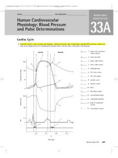
6 During physical work, 5x more 4) Isoionia: Isoionic environment (including perfusion fluid) Balance between: Calcium and potassium During Ca abundance rigor During K abundance ' inhibition 5) pH: acidosis inhibition of the heart activity heart stops in diastole alkalosis heart stops in systole rigor The Cardiac Cycle ' the period from the end of one cardiac systole to the end of the next heart contraction. 1) Electrical cycle depolarisation/repolarisation 2) Mechanical cycle contraction/relaxation of cardiac muscle Periods of the cardiac cycle 1) Filling of the atria during diastole = venous return Mechanisms of the filling: a) Vis a tergo residual energy from the left ventricle b) Negative intrathoracic (interpleural) pressure: Quiet breathing: ' expiration: Ppl = ' mm Hg (relative to atmospheric) ' inspiration: '6 mm Hg during = thoracic pump.
7 Pressure is transmitted to the great veins and atria aids venous return. The movement of diaphragm rise of Pabd. c) Shifting down of the A'V ring by contraction of ventricles vis a fronte . d) Gravity from head and vessels above cardiac level e) Muscular pump Filling of the ventricles Accumulation of blood in the atria elevated atrial pressure ' decrease of ventricular pressure to diastolic value (near ) pressure in the atria push open the A'V valves blood flows into the ventricles 1) Period of rapid filling (first 1/3 of the diastolic time) 2) Period of slow filling diastasis (next 1/3) 3) Atrial systoly (last 1/3) + 20'30 % of the filling of the ventricles Ventricular systole 1) Period of isovolumic.
8 ' isometric contraction At the start of ventricular contraction, the ventricular pressure rises causing the A'V valves to close. Also semilunar valves are closed during first about s until the pressures in LV and RV and RV exceed the pressures in the aorta and pulmonary artery opening of the semilunar valves 2) Period of ventricular ejection a) Phase of the rapid ejection (1/2 of V is ejected in the first of the ventricular systole) b) Phase of the slow ejection (remaining of V'during next 2/4 of the ventricular systole) 3) Protodiastole (last of the ventricular systole) The ventricular pressure ralls to a value below that in aorta, closing of the semilunar valves early diastole.
9 Ventricular diastole 1) Period of isovolumic (isometric) relaxation the valves are closed,V pressures continues to drop 2) Filling of the ventricles: Period of the rapid ventricular filling when the ventricular pressure falls below atrial pressure and the AV valves open ..a new cycle. HR = 72/min 1 cycle lasts s. Length of Systole/Diastole HR 65/min: S ' s D ' s 1 : 2 HR 200/min: S ' s D ' s 1 : 1 The duration of systole is more fixed. Tachycardia is accompanied mainly by shortening of the diastole if more than 180/min insufficient filling critical frequency (HR) for adults. Functions of the valves The AV valves prevent backflow of blood from V to A during systole The semilunar valves prevent backflow from the aorta and pulmonary artery to V during diastole.
10 All valves close and open passively by pressure gradient. The Electrical Activity of the Heart Resting membrane potential (RMP): myocardial fibers approximately 90 mV SA node: '55 to 60 mV Conductive tissue: ' 90 to 100 mV +++++++++ ' ' ' ' ' ' ' ' ' ' ' ' ' ' ' ' ' ' +++++++++ RMP depends on differences in concentration of K+ Ki+150 mmol/l; Ke+ ' 5 mmol/l = 30 x RMP don t allow to K+ to equalize concentrations. Nai+ = 5'10 mmol/l; Nae+ = 140 mmol/l Depolarization: Firing level 65 mV Initial is due to an increase in Na+ permeability (through fast Na+ channels) Following a slower increase in Ca2+ permeability (through slow Ca2+ channels) plateau (!)