Example: dental hygienist
Streptavidin MicroBeads - miltenyibiotec.com
fluorochrome conjugated streptavidin for flow cytometric analysis. (Optional) Propidium iodide (PI) or 7-AAD for flow cytometric exclusion of dead cells. (Optional) Pre-Separation Filters (#130-041-407) to remove cell clumps. (Optional) Dead Cell Removal Kit (#130-090-101) for the
Tags:
Information
Domain:
Source:
Link to this page:
Documents from same domain

Isolation of mononuclear cells from human peripheral blood ...
www.miltenyibiotec.comThe peripheral blood or buffy coat should not be older than 8 hours and supplemented with anticoagulants (e.g. heparin, EDTA, citrate, ACD-A, or citrate phosphate dextrose (CPD)). 1. Dilute cells with 2–4× the volume of buffer. Note: The more diluted the blood sample, the better the purity of the

Vio® Dyes technical specifications - Miltenyi Biotec
www.miltenyibiotec.comHuman peripheral blood mononuclear cells (PBMC) and mouse splenocyte (MS) cells stained with VioBlue® exhibit excellent discrimination between positively and negatively stained populations over a wide range of antibody specificities (figure 3). Furthermore, VioBlue provides a superior alternative

Dead Cell Removal Kit - Miltenyi Biotec
www.miltenyibiotec.com4. Use the wash program Clean. 5. (Optional) For elution of dead cells (labeled cells), press Pos. Cells will be eluted in tube containing 1× Binding Buffer in row C. At this step, remaining volume of 1× Binding Buffer in the tube will be 0.5mL. To discard dead cells, press Waste. 3. Example for elimination of dead cells from

T Cell TransAct™ human - Miltenyi Biotec
www.miltenyibiotec.com2.1 Sample preparation ... by flow cytometry or fluorescence microscopy. nless therwse spec⁶call ncate, all lten tec pructs an serces page 3/3 140-004-952.05 are r research use nl an nt r agnstc r therapeutc use. 3. Refer to Examples of T cell activation and expansion ... fluorescence. Negative control

MS Columns
www.miltenyibiotec.comsmall, superparamagnetic particles, a high-gradient magnetic field is required to retain the labeled cells. MS Columns contain an optimized matrix to generate this strong magnetic field when ... from material defects in workmanship and materials and to conform substantially

MACS® Cell Separation - Miltenyi Biotec
www.miltenyibiotec.com• Minimal cell labeling with nano-sized MicroBeads ensures preservation of cellular integrity and characteristics • Used in over 55,000 clinical cellular treatments to date Figure 1: It only takes three easy steps to get viable cells with high yield and purity from your sample. Watch how to isolate cells in three easy steps at
Related documents

Introduction to Flow Cytometry - Boston University
www.bu.eduAll flow cytometric measurements can be made simultaneously on a single cell. TF 10 Particles must be in single-cell suspension before flow cytometric analysis. TF. Introduction to Flow Cytometry: A Learning Guide 4. 5 2 Fluidics The purpose of the fluidics system is to transport particles in a fluid stream to the laser
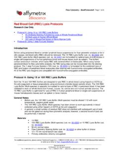
Red Blood Cell (RBC) Lysis Protocols - tools.thermofisher.com
tools.thermofisher.comperipheral blood for flow cytometric analysis, the RBC lysing step can be incorporated into the staining protocol. The 1-step Fix/Lyse Solution (10X) (cat. no. 00-5333) is formulated for the combined lysis of RBC and fixation of peripheral blood leukocytes after staining with fluorochrome-conjugated antibodies.
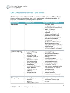
CAP Accreditation Checklists 2021 Edition
documents.cap.org• Rare event flow cytometric assays Forensic Drug Testing •Drug Testing –Hair • Drug Testing – Meconium • for different specimen typesDrug Testing – Nails • Drug Testing – Oral fluid • Drug Testing – Umbilical Cord • Drug Testing – Urine • Drug Testing – Urine Screen Only • Drug Testing – Whole Blood

The Annexin V Apoptosis Assay - University of Kansas ...
www.kumc.edu∞ 1994 Koopman et al. developed a flow cytometric assay using Annexin V conjugated to FITC to measure Annexin V binding to apoptotic cells. The authors stained control and serum starved cells with ethidium bromide and Annexin V-FITC. Binding of Annexin V correlated with other

BD FACS Lysing Solution - bdbiosciences.com
www.bdbiosciences.comdark until flow cytometric analysis. Analyze the stained cells within the time limit specified in the appropriate IFU. Vortex the cells thoroughly at low speed to reduce aggregation before acquiring. 9. RESULTS The following representative data was obtained with peripheral blood samples treated with BD FACS lysing solution on a

Basic Hematology
www.hematology.orga) This is now determined directly by electrical or flow cytometric methods. b) it is always calculated from the spun hematocrit. c) it is determined by light microscopy.

Isolectin IB4 Conjugates - Thermo Fisher Scientific
assets.thermofisher.comcells.4 It has also been particularly valuable as a histochemical and flow cytometric probe for specifically labeling endothelial cells in a number of species.5,6 Isolectin IB 4 has been used ef-fectively for tracing central and peripheral neuronal pathways following local injections,7,8 as

Kit Manual - bdbiosciences.com
www.bdbiosciences.comFor Research Use Only. Not for use in diagnostic or therapeutic procedures. 6 www.bdbiosciences.com Kit components: Fixation/Permeabilization solution (125 mL) BD Perm/Wash™ Buffer, 10 × concentrate containing FBS* and saponin (dilute 1:10 in distilled H