Transcription of Autophagy - Abcam
1 AutophagyTools for cell death series 23 ContentsIntroduction to cell death ..3 Autophagy ..3 -Types of Autophagy ..4 Macroautophagy ..4 Microautophagy ..4 Chaperone mediated Autophagy ..4 Autophagy : cellular and molecular mechanisms ..6 -Induction and phagophore formation ..8 -Phagophore elongation and autophagosome formation ..10 -Fusion, degradation and recycling ..12 - Autophagy regulation ..14 Autophagy and disease -Cancer ..14 -Neurodegenerative diseases ..15 -Cardiovascular diseases ..15 -Infectious disease ..16 Autophagy detection -Markers of Autophagy ..17 -Visual identification ..19 -ATG8/LC3 detection ..20 Western Blotting ..20 Turnover of LC3 II/ATG8 PE ..22 Luminescence based assays ..25 GFP ATG8/LC3 detection ..25 Fluorescence microscopy ..26 Tandem mRFP/mcherry GFP fluorescence microscopy ..28 Flow and multispectral imaging cytometry.
2 29 Immunohistochemistry ..31 -SQSTM1/p62 and related LC3 binding protein detection ..32 -TOR, AMPK and ATG1/ULK1 detection ..33 -Additional Autophagy related markers ..34 ATG9 ..34 ATG14 ..34 ATG18 ..35 BECLIN1 .. 35 DRAM1 ..35 General considerations for Autophagy detection ..36 References ..374 Introduction to cell deathCell death happens when a biological cell fails to maintain essential life functions . This may be the result of the natural process of development or protection against cellular defects, of normal cells replacement, or factors such as disease or localized injury .Cell death can be classified according to its morphological appearance (such as apoptotic or necrotic), enzymological criteria (with or without involvement of distinct proteases), functional aspects (programmed or accidental) or immunological characteristics (immunogenic or non immunogenic).
3 In this guide, we want to examine the role of Autophagy , a self degradation process that has been historically linked to non apoptotic cell death .5 AutophagyAutophagy (from the Greek auto self and phagein to eat) is a general term for processes by which dysfunctional cellular components are degraded inside the cell through the action of lysosomes . Components of the cytoplasm, including organelles, are sequestered and moved into the lumen of lysosomes/vacuoles where they are broken down into basic components and returned to the cytosol for reuse .Under physiological conditions, Autophagy has a number of vital roles such as maintenance of the amino acid pool during starvation, preimplantation development, clearance of intracellular microbes, and regulation of innate and adaptive immunity1 . Autophagy has been widely implicated in many pathophysiological processes such as cancer, metabolic and neurodegenerative disorders as well as cardiovascular and pulmonary diseases.
4 It also has an important role in aging and exercise2 .One of the characteristic features of Autophagy is its dynamic regulation; cellular autophagic activity is usually low under basal conditions, but there are a number of stimuli that can lead to its upregulation . Nutrient starvation is the most well known inducer of Autophagy , but other biological stress stimuli such as hypoxia or energy depletion, hormonal stimulation or pharmaceutical agents can increase Autophagy in the cells as well . Autophagy is a common morphological feature in dying cells, and therefore it was presumed for a long time to be a cell death pathway . Recent investigations show that one of its major functions is to work on keeping cells alive under stressful conditions that could otherwise lead to cell death .Types of autophagyThere are currently three types of Autophagy in mammalian cells: macroautophagy, microautophagy and chaperone mediated Autophagy , macroautophagy being the most extensively studied type3.
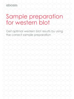
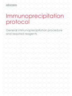
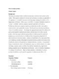
5 MacroautophagyMacroautophagy is the main autophagic pathway, generally referred to as Autophagy . It is characterized by the delivery of cytoplasmic cargo to the lysosome through an intermediary double membrane bound vesicle, known as autophagosome, which fuses with the lysosome to form an autolysosome .Throughout this guide we will review the cellular and molecular mechanism involved in macroautophagy (referred to as Autophagy from now on), as well as methods and assays which can be used to monitor Autophagy . MicroautophagyMicroautophagy, on the other hand, involves the direct engulfment of cytoplasmic cargo into the lysosome through invagination of the lysosomal membrane . Microautophagy is important in the maintenance of organellar size, membrane homeostasis and cell survival under nitrogen restriction4 .6 Chaperone mediated autophagyChaperone mediated Autophagy (CMA) involves the direct translocation of cytoplasmic proteins across the lysosomal membrane in a complex with chaperone proteins that are recognized by the lysosomal membrane receptor LAMP 2A (lysosomal associated membrane protein 2A), resulting in their unfolding and degradation.
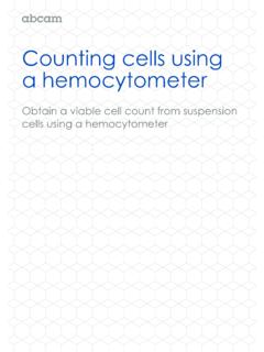
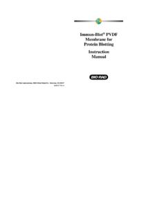
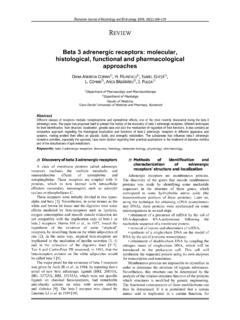
6 IsolationmembraneProteinKFERQ motifHsc70chaperoneLamp2 AChaperoneMediatedAutophagyCytosolPhagop horeAutophagosomeMacro-autophagyMicroaut ophagyLysosomeFigure 1. Types of Autophagy7 Autophagy : Cellular and molecular mechanismsAutophagy is a tightly regulated pathway which at basal level has an important housekeeping role allowing cells to eliminate damaged or harmful components through catabolism and recycling to maintain nutrient and energy homeostasis . Autophagy is also a major protective mechanism which allows cells to survive in response to multiple stress conditions such as nutrient or growth factor deprivation, hypoxia, reactive oxygen species, DNA damage or intracellular pathogens5 .The process of Autophagy begins with an expanding membrane structure known as phagophore which enwraps portions of the cytoplasm . This leads to the formation of a double membrane sequestering vesicle, called autophagosome.
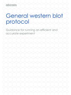
7 Autophagosomes subsequently fuse with lysosomes, releasing their inner compartment to the lysosomal lumen . The inner membrane part of the autophagosome, together with the enclosed cargo, is degraded, and the resulting macromolecules are released into the cytosol through lysosomal membrane permeases for recycling6 .IsolationmembraneInduction andphagophoreformationElongation andautophagosomeformationLysosomefusionD egradationand recyclingStarvationHypoxiaROSI nfectionChemotherapyOthersPhagophoreAuto phagosomeAutolysosomeRecyclingDegradatio nLysosomeFigure 2. Autophagy process overview8 Induction and phagophore formationThe molecular mechanism of Autophagy involves several conserved Atg ( Autophagy related) proteins . In response to various stimuli (nutrient starvation, hypoxia, ROS, infection, DNA damage and others) a unique flat membrane (phagophore) is formed . Two protein complexes are involved in this initiation step: one complex contains the class III PI3 K/Vps34, Atg6/Beclin1, Atg14, and Vps15/p150, and the other complex includes a serine/threonine kinase Atg1/ULK1 which is an essential positive regulator of autophagosome formation.
8 When nutrients are abundant, binding of the ULK1 complex by the mammalian target of rapamycin (mTOR) complex 1 (mTORC1) inhibits Autophagy . The mTORC1 is an important regulator of cell growth and metabolism . It is composed of five subunits that include Raptor, which binds ULK1, and mTOR, a serine threonine kinase . By phosphorylating ULK1, mTOR inhibits Autophagy initiation . In starvation, mTORC1 dissociates from the ULK1 complex, freeing it to trigger autophagosome nucleation and elongation . The kinase activity of Atg1 requires the function of two other Autophagy proteins, Atg13 or Atg8 and Atg177 .Figure 3. Induction and phagophore formation9 StarvationHypoxiaROSI nfectionChemotherapyOthersAutophagosomeC lass III PI 3-K complexLC3 conjugation systemAtg12 conjugation systemAtg/ULK1 complexLAMP2 Atg14 LBeclin 1hVps34 Ulk1/2 Atg101 Atg13 FIP200hVps15 Ambra1 Atg22 Rab7 Atg9 Atg2 Atg5 Atg12 Atg4 Atg16 Atg16 UVRAGR ubiconSNARESHOPSVMP1 WIPI1/2 Atg5 PhagophoreformationInductionElongation andautophagosome formationLysosomefusionDegradationand recyclingAtg7LC3-IILC3-ILC3 Atg5 Atg12 Atg12 PEPE10 Phagophore elongation and autophagosome formationThe elongation of the phagophore results in the formation of autophagosome, which is typically a double membraned organelle.
9 This step is a simple sequestration, and no degradation occurs . Phagophore elongation and autophagosome completion requires two ubiquitin like conjugation pathways, both catalyzed by Atg7 .The first leads to the conjugation of Atg5 Atg12 which then form a multimeric complex with Atg16L . The Atg5 Atg12 Atg16L complex associates with the outer membrane of the extending phagophore3,8 . The association of Atg5 Atg12 Atg16L complexes is thought to induce curvature into the growing phagophore through asymmetric recruitment of processed LC3B II . Atg5 Atg12 conjugation is not dependent on activation of Autophagy and once the autophagosome is formed, Atg5 Atg12 Atg16L dissociates from the membrane, making conjugated Atg5 Atg12 a relatively insufficient marker of autophagy9 .The second ubiquitin like system involved in autophagosome formation results in the processing of microtubule associated protein light chain 3 (LC3B), which is encoded by the principal mammalian homologue of the yeast Atg8.
10 Upon Autophagy induction LC3B, expressed in most cell types as a full length cytosolic protein, is proteolytically cleaved by Atg4 to generate LC3B I . LC3B I is activated by Atg7 and then transferred to Atg3 followed by conjugation to phosphatidylethanolamine (PE) to generate processed LC3B II .Processed LC3B II is recruited onto the growing phagophore and its integration is dependent on Atg5 Atg12 . Unlike Atg5 Atg12 Atg16L, LC3B II is found on both the inner and the outer surfaces of the autophagosome, where it is required for the expansion and completion of the autophagic membrane and plays a role in selecting cargo for degradation as well as in the fusion of the autophagosome with the lysosome . LC3B II has been proposed to act as a receptor for selective substrate, p62/SQSTM110 . During Autophagy the synthesis and processing of LC3 is increased . Recent research indicates that the deacetylation and cytosolic translocation of a nuclear pool of LC3 is required for its lipidation with PE during starvation induced autophagy11.