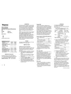
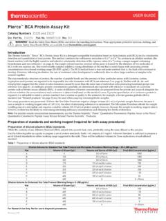
Transcription of INSTRUCTIONS Micro BCA Protein Assay Kit
1 INSTRUCTIONS Pierce Biotechnology PO Box 117 (815) 968-0747 3747 N. Meridian Road Rockford, lL 61105 USA (815) 968-7316 fax Number Description 23235 Micro BCA Protein Assay Kit, sufficient reagents for 480 tube assays or 3200 microplate assays Kit Contents: Micro BCA Reagent A (MA), 240mL Micro BCA Reagent B (MB), 240mL Micro BCA Reagent C (MC), 12mL bovine serum albumin Standard Ampules, 2mg/mL, 10 1mL ampules containing bovine serum albumin (BSA) at in a solution of saline and sodium azide Storage: Upon receipt store product at room temperature. Product shipped at ambient temperature. Note: If either Reagent MA or Reagent MB precipitates upon shipping in cold weather or during long-term storage, dissolve precipitates by gently warming and stirring solutions.
2 Discard any reagent that shows discoloration or evidence of microbial contamination. Table of Contents Introduction .. 1 Preparation of Standards and Working Reagent .. 2 Test Tube Procedure (linear working range of g/mL) .. 3 Microplate Procedure (linear working range of 2-40 g/mL) .. 3 Troubleshooting .. 3 Additional Information .. 4 Related Thermo Scientific Products .. 5 References .. 5 Introduction The Thermo Scientific Micro BCA Protein Assay Kit is a detergent-compatible bicinchoninic acid formulation for the colorimetric detection and quantitation of total Protein . An adaptation of the Thermo Scientific BCA Protein Assay Kit (Product No.)
3 23225), the Micro BCA Kit has been optimized for use with dilute Protein samples ( g/mL). The unique, patented method uses bicinchoninic acid (BCA) as the detection reagent for Cu+1, which is formed when Cu+2 is reduced by Protein in an alkaline A purple-colored reaction product is formed by the chelation of two molecules of BCA with one cuprous ion (Cu+1). This water-soluble complex exhibits a strong absorbance at 562nm that is linear with increasing Protein concentrations. The macromolecular structure of Protein , the number of peptide bonds and the presence of four amino acids (cysteine, cystine, tryptophan and tyrosine) are reported to be responsible for color formation with Studies with di-, tri- and tetrapeptides suggest that the extent of color formation is caused by more than the mere sum of individual color-producing functional The Micro BCA Protein Assay Kit uses concentrated reagents and a protocol that utilizes an extended incubation time at an elevated temperature (60 C, Test Tube Procedure only).
4 The result is an extremely sensitive colorimetric Protein Assay in a test tube or microplate Assay format. Note: For peptide sample concentration measurements, use the Thermo Scientific Pierce Quantitative Fluorometric Peptide Assay or the Pierce Quantitative Colorimetric Peptide Assay (see Related Thermo Scientific Products). 23235 Micro BCA Protein Assay Kit Pierce Biotechnology PO Box 117 (815) 968-0747 3747 N. Meridian Road Rockford, lL 61105 USA (815) 968-7316 fax 2 Preparation of Standards and Working Reagent A. Preparation of Diluted albumin (BSA) Standards Use Table 1 as a guide to prepare a set of Protein standards.
5 Dilute the contents of one albumin (BSA) Standard ampule into several clean vials, preferably using a diluent that is similar to the sample buffer. Each 1mL ampule of albumin Standard is sufficient to prepare a set of diluted standards such that three replicates of each dilution may be included in the Test Tube Procedure. Table 1. Preparation of Diluted albumin (BSA) Standards Vial Volume of Diluent Volume and Source of BSA Final BSA Concentration A of Stock 200 g/mL B of vial A dilution 40 g/mL C of vial B dilution 20 g/mL D of vial C dilution 10 g/mL E of vial D dilution 5 g/mL F of vial E dilution g/mL G of vial F dilution 1 g/mL H of vial G dilution g/mL I 0 0 g/mL = Blank B.
6 Preparation of the Micro BCA Working Reagent (WR) 1. Use the following formula to determine the total volume of WR required: (# standards + # unknowns) (# replicates) (volume of WR per sample) = total volume WR required Example: for the standard Test Tube Procedure with 3 unknowns and 2 replicates of each sample: (9 standards + 3 unknowns) (2 replicates) (1mL) = 24mL WR required (round up to 25mL) Note: 1mL of the WR is required for each sample in the Test Tube Procedure, while only 150 L of WR is required for each sample in the Microplate Procedure. 2. Prepare WR by mixing 25 parts of Micro BCA Reagent MA and 24 parts Reagent MB with 1 part of Reagent MC (25:24:1, Reagent MA:MB:MC).
7 For the above example, combine of Reagent MA and Reagent MB with of Reagent MC. Note: When Reagent MC is initially added to Reagents MA and MB, turbidity occurs that quickly disappears upon mixing to yield a clear-green solution. Prepare sufficient volume of WR based on the number of samples to be assayed. The WR is stable for one day when stored in a closed container at room temperature (RT). It is not necessary to protect the solution from light. Procedure Summary (Test Tube Procedure) Pierce Biotechnology PO Box 117 (815) 968-0747 3747 N. Meridian Road Rockford, lL 61105 USA (815) 968-7316 fax 3 Test Tube Procedure (linear working range of g/mL) 1.
8 Pipette of each standard and unknown sample replicate into appropriately labeled test tubes. 2. Add of the WR to each tube and mix well. 3. Cover tubes and incubate at 60 C in a water bath for 1 hour. 4. Cool all tubes to room temperature (RT). 5. With the spectrophotometer set to 562nm, zero the instrument on a cuvette filled only with water. Subsequently, measure the absorbance of all the samples within 10 minutes. Note: Color development continues even after cooling to RT. However, the rate of development at RT is sufficient low that no significant error is introduced if all absorbance measurements are made within a 10-minute period.
9 6. Subtract the average 562nm absorbance reading of the Blank standard replicates from the 562nm reading of all other individual standard and unknown sample replicates. 7. Prepare a standard curve by plotting the average Blank-corrected 562nm reading for each BSA standard vs. its concentration in g/mL. Use the standard curve to determine the Protein concentration of each unknown sample. Microplate Procedure (linear working range of 2-40 g/mL) 1. Pipette 150 L of each standard or unknown sample replicate into a microplate well (Product No. 15041). 2. Add 150 L of the WR to each well and mix plate thoroughly on a plate shaker for 30 seconds.
10 3. Cover plate using Sealing Tape for 96-Well Plates (Product No. 15036) and incubate at 37 C for 2 hours. Note: Limit incubations of microplate to less than or equal to 37 C, otherwise high background and aberrant color development may result. Most polystyrene Assay plates deform, leach, and become cloudy at 60 C. 4. Cool plate to room temperature (RT). 5. Measure the absorbance at or near 562nm on a plate reader. 6. Subtract the average 562nm absorbance reading of the Blank standard replicates from the 562nm reading of all other individual standard and unknown sample replicates. 7. Prepare a standard curve by plotting the average Blank-corrected 562nm reading for each BSA standard vs.