Transcription of 15.2 ULTRAVIOLET–VISIBLE SPECTROSCOPY
1 684 CHAPTER 15 DIENES, RESONANCE, AND visible SPECTROSCOPYThe IR and NMR spectra of conjugated dienes are very similar to the spectra of ordinaryalkenes. However, another type of SPECTROSCOPY can be used to analyze and identify organiccompounds containing conjugated p-electron systems. In this type of SPECTROSCOPY , called ul-traviolet visible SPECTROSCOPY ,the absorption of radiation in the ultraviolet or visible regionof the spectrum is recorded as a function of wavelength. The part of the ultraviolet spectrumof greatest interest to organic chemists is the near ultraviolet (wavelength range 200X 10_9to 400X 10_9m). visible light, as the name implies, is electromagnetic radiation visible to thehuman eye (wavelengths from 400X 10_9to 750X 10_9m).
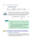
2 Because there is a commonphysical basis for the absorption of both ultraviolet and visible radiation by chemical com-pounds, both ultraviolet and visible SPECTROSCOPY are considered together as one type of spec-troscopy, often called simply UV vis The UV Vis SpectrumLike any other absorption spectrum, the UV vis spectrumof a substance is the graph of radi-ation absorption by the substance versus the wavelength of the radiation. The instrument usedto measure a UV vis spectrum is called a UV vis for the fact thatit is designed to operate in a different part of the electromagnetic spectrum, it is conceptuallymuch like any other absorption spectrometer (Fig. , p. 539).A typical UV spectrum, that of 2-methyl-1,3-butadiene (isoprene), is shown in Fig.
3 Isoprene does not absorb visible light, only the ultraviolet region of the spectrum isshown. On the horizontal axis of the UV spectrum is plotted the wavelength lof the UV SPECTROSCOPY , the conventional unit of wavelength is the nanometer(abbreviated nm).One nanometerequals 10_9meter. (In older literature, the term millimicron,abbreviated mm,was used; a millimicron is the same as a nanometer.) The relationship between the energy ofthe electromagnetic radiation and its frequency or wavelength should be reviewed again(Sec. ). = nmCH3CH2 CCH2 CHwavelength (l), nmFigure visible spectrum of isoprene in methanol. The lmax(red) is the wavelength at which theabsorption maximum occurs; for isoprene, the lmaxis 12/9/08 12:22 PM Page ultraviolet visible SPECTROSCOPY685 The vertical axis of a UV spectrum shows the absorbance.
4 (Absorbance is sometimes calledoptical density,abbreviated OD.) The absorbance is a measure of the amount of radiant energyabsorbed. Suppose the radiation entering a sample has intensity I0, and the light emerging fromthe sample has intensity I. The absorbance Ais defined as the logarithm of the ratio I0 I:A= log (I0 I)( )According to Equation , then, the more radiant energy is absorbed, the larger is the ratioI0 I, and the greater the the UV vis spectra used in this text, absorbance increases from the bottom to the topof the spectrum. Therefore, absorption maxima occur as high points or peaks in the spec-trum. Notice the difference in how UV vis and IR spectra are presented. (Absorptions inIR spectra increase from top to bottom because IR spectra are conventionally presented asplots of transmittance,or percentage of light transmitted.)
5 In the UV vis spectrum shownin Fig. , the absorbance maximum occurs at a wavelength of nm. The wavelengthat the maximum of an absorption peak is called the lmax(pronounced lambda-max ).Some compounds have several absorption peaks and a corresponding number of lmaxval-ues. Absorption peaks in the UV vis spectra of compounds in solution are generally quitebroad. That is, peak widths span a considerable range of wavelength, typically 50 nm ormore. (The reason is discussed in Further Exploration in the Study Guide.)The absorbance at a given wavelength depends on the number of molecules in the lightpath. If a sample is contained in a vessel with a thickness along the light path of lcm, and theabsorbing compound is present at a concentration of cmoles per liter, then the absorbance isproportional to the product elc( )This equation is called the Beer Lambert lawor simply Beer s constant of propor-tionality eis called the molar extinction coefficientor molar units of eareLmol_1cm_1, or M_1cm_1; these units are sometimes omitted when values of eare absorption in a given spectrum has a unique extinction coefficient that depends on wave-length, solvent, and temperature.
6 The larger is e, the greater is the light absorption at a givenconcentration cand path length l. For example, the extinction coefficient of isoprene ( ) at its lmaxof nm is 10,750 M_1cm_1in methanol solvent at 25 C; its extinctioncoefficient in alkane solvents is nearly twice as coefficients of 104 105M_1cm_1are common for molecules with conjugatedp-electron systems. This means that strong absorptions can be obtained from very dilute so-lutions solutions with concentrations on the order of 10_4 10_6 Mwith a typical pathlength of 1 cm. Because of its intrinsic sensitivity and its relatively simple instrumentation,UV vis SPECTROSCOPY was one of the earliest forms of SPECTROSCOPY to be used routinely What is the energy of light (in kJ mol_1or kcal mol_1) with a wavelength of(a)450 nm?
7 (b) 250 nm? (a)What percent of the incident radiation is transmitted by a sample when its absorbance When its absorbance is 0?(b) What is the absorbance of a sample that transmits one-half of the incident radiationintensity? A thin piece of red glass held up to white light appears brighter to the eye than a piece of thesame glass that is twice as thick. Which piece has the greater absorbance?PROBLEMSF urther Exploration on 12/9/08 12:22 PM Page 685686 CHAPTER 15 DIENES, RESONANCE, AND AROMATICITYthe laboratory; adequate spectra could be obtained on even the most primitive spectrome-ters. UV vis SPECTROSCOPY remains a very important method for quantitative UV vis spectra are presented in abbreviated form by citing the lmaxvalues of theirprincipal peaks, the solvent used, and the extinction coefficients.
8 For example, the spectrum inFig. is summarized as follows:lmax(CH3OH)= nm (e= 10,750)orlmax(CH3OH)= nm (log e= )B. Physical Basis of UV Vis SpectroscopyWhat determines whether an organic compound will absorb UV or visible radiation? ultraviolet and/or visible radiation is absorbed by the pelectrons and, in some cases, by theunshared electron pairs in organic compounds. For this reason, UV vis spectra are sometimescalled electronic spectra. (The electrons of sbonds absorb at much shorter wavelengths, inthe far ultraviolet .) Absorptions by compounds containing only single bonds and unsharedelectron pairs are generally quite weak (that is, their extinction coefficients are small).
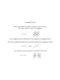
9 How-ever, intense absorption of UV and visible radiation occurs when a compound contains pelec-trons. The simplest hydrocarbon containing pelectrons, ethylene, absorbs UV radiation atlmax=165 nm (e=15,000). Although this is a strong absorption, the lmaxof ethylene andother simple alkenes is below the usual working wavelength range of most conventionalUV vis spectrophotometers; the lower end of this range is about 200 nm. However, moleculeswith conjugateddouble or triple bonds (for example, isoprene, Fig. ) have lmaxvaluesgreater than 200 nm. Therefore, UV vis SPECTROSCOPY is especially useful for the diagnosis ofconjugated double or triple structural feature of a molecule responsible for its UV vis absorption is called a chro-mophore, from Greek words meaning to bear color.
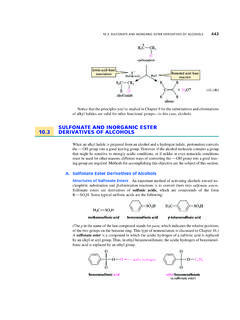
10 Thus, the chromophore in isoprene(Fig. ) is the system of conjugated double bonds. Because many important compounds donot contain conjugated double bonds or other chromophores, UV vis SPECTROSCOPY has lim-ited utility in structure determination compared with NMR and IR SPECTROSCOPY . However, thetechnique is widely used for quantitative analysis in both chemistry and biology; and, whenthe double bonds are not conjugated;no lmax above 200 nmCH2H2 CCHCH2CH2 CHAAthe double bonds are conjugated; this compound has a lmax above 200 nm: lmax = 227 nm (e = 14,200)CH3H"H3 CCH"CH"CH"C%((%% (a)From the extinction coefficient of isoprene (10,750 M_1cm_1) and its observed ab-sorbance at nm (Fig.)))