Transcription of Exercise Physiology: Cardiovascular System - Colby College
1 1/9/13 Philip Melillo - Exercise physiology Cardiovascular System1 | About Us | Contact UsExercise physiology : Cardiovascular SystemIntroductionAnatomy of the HeartChambers, Values, and Blood Flow within HeartCardiac FunctionThe Blood Vessels and Circulation (The Vasculature) Formula Recap (Blood Pressure, Stroke Volume and CardiacOutput)References IntroductionI became inspired to write this article when I realized that so many people, even those you Exercise regularly and participate inendurance sports, did not fully understand the basics of Cardiovascular Exercise physiology . My goal here is to keep it simple, yetestablish a foundation so when I write about the acute and chronic effect that endurance Exercise has on the cardiovascularsystem, you will have a better understanding of how and why the body reacts in the way it does.
2 Also, you will be in a betterposition to optimize your training to meet your study of the Cardiovascular Exercise physiology is one of the significant disciplines of Exercise physiology . It examines howoxygen and other nutrients are transported by Cardiovascular System and used by the muscles during primary purpose of the System is to deliver nutrients to and remove metabolic waste products from tissues. It performs thefollowing functions [1]:Transports deoxygenated blood from the heart to the lungs and oxygenated blood from the lungs to the heartTransports oxygenated blood from the heart to tissues and deoxygenated blood from the tissues to the heartDistributes nutrients to cells ( glucose)Removes metabolic wastes ( lactate)Regulates pH to control acidosis and alkalosisTransports hormones and enzymes to regulate physiological functionMaintains fluid balance to prevent dehydrationMaintains body temperature by absorbing and redistributing heat Anatomy of the HeartThe heart is a muscular organ that has two interconnected but separate pumps.
3 Each pump has two chambers an atrium (upper)and a ventricle (lower). The right side of the heart pumps collects deoxygenated blood from the periphery and pumps it through thelungs (pulmonary circuit). The left side collects blood from the lungs andpumps it throughout the body (systemic circuit).The sulcli, the external deep grooves of the heart, define the four chambers right atrium (RA), left atrium (LA), right ventricle (RV),and left ventricle (LV) [2]. The coronary sulcus separates the atria from the ventricles. The interventricular sulcus separates the rightventricle and the left heart has a base and an apex. The base consists mainly of the LA and RA, and parts of the proximal portion of the large is located above and close to the right sternal border at the level of the second and third ribs.
4 The apex of the heart is locatedbelow the base at the level of the fifth intercostal Melillo - Exercise physiology Cardiovascular System2 heart is covered by a double walled loose sitting membrane called the pericardium. The interior lining of the heart is called theepicardium. The cardiac muscle, the thickest layer of tissue in the heart,is called the myocardium. The myocardium contains a network ofconnective tissue fibers called the fibrous skeleton which separates theatria from the ventricles and provides support for the myocardium and thevalues of the , Values, and Blood Flow within the HeartAs I stated earlier, the heart has four chambers right atrium (RA), leftatrium (LA), right ventricle (RV), and left ventricle ( LV ).
5 The RA and LAfunction primarily as blood reservoirs, delivering blood into the RV andLV, respectively. The RV supplies the main force for moving bloodthrough the pulmonary circuit. The left LV supplies the main force formoving blood through the systemic heart has four values whose function is to keep the blood flowing inone direction. The atrioventricular (AV) valves separate the atria from theventricles. The right AV valve has three cusps and is called the tricuspidvalue. It controls the blood flow from the RA to the RV. The left AV hastwo cusps and is called the mitral or bicuspid value. The mitral valuecontrols blood flow between the LA and are two semilunar valves pulmonic and aorta.
6 The pulmonicvalve lies between the RV and the pulmonary artery. The aortic valve lies between the LV and the aorta. They separate theventricles from the pulmonary artery and aorta, respectively. Both semilunar valves prevent the backflow of blood to the flow though the heart is accomplished by the following sequence of events starting with the return of blood from the Deoxygenated blood flows into the RA through the superior and inferior vena cavae, the coronary sinus, and anterior cardiacveins2. RA contracts, and blood moves through the tricuspid value into the RV3. The RV contracts, the tricuspid closes, and blood flows through the pulmonic value into the pulmonary arteries4.
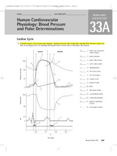
7 Blood enters the alveolar capillaries where oxygen is absorbed and carbon dioxide is removed5. Blood flows back to the LA through the pulmonary veins6. The LA contracts, and blood flows through the mitral valve and into the LV7. The LV contracts, the mitral value closes, the blood flows through though the aortic value into the aorta where it's thendistributed to the coronary circulation and the systemic circulation [3]Figure 1 depicts the heart's anatomy including the blood flow. Please excuse my drawing as you can see, illustrations is not mystrong point. I hope you at least get the Heart's Electrical Conduction SystemCardiac muscle has unique properties that allow it to contract without an external nervous System .
8 The electrical stimulus for themechanical contraction is provided by a specialized electrical conduction System . This System consists of the sinoatrial (SA) node,atrioventrical (AV) node, AV bundle, right and left branches, and the Purkinje into the detail of how this System functions is outside the scope of this article. For now just know that the SA node normallycontrols the rhythm of the electrical stimulation of the heart and ultimately the heart's contraction patterns and that this is influencedby the brain's Cardiovascular center (medulla) that transits signals to the heart through two components of the autonomic nervoussystem the sympathetic nervous System and the parasympathetic nervous System .
9 Stimulation of the sympathetic nervesaccelerates firing of the SA node, causing the heart to beat faster. Simulation of the parasympathetic nervous System slows the ratethat the SA node fires, thus slowing the heart rate. In another article I'll explain the effect that Exercise and stress has on thesenervous systems and thus heart FunctionThe topics of heart rate, stroke volume, and cardiac output are important as they are key elements when I discuss the effects ofendurance Exercise on the cardiac function in other RateHeart rate (HR) is number of heart beats per minute. The average normal resting HR is between 60 and 80 beats per minute (bpm).
10 Resting HR in women is usually 10 bpm higher then men. Children have higher heart rates than adults elderly people have lowerheart rates and fit people in the same age group and gender have lower resting HR than unfit people. Stroke VolumeStroke volume (SV) is the amount of blood ejected from the left ventricle (LV) in a single contraction measured in milliliters (mL). Atrest, SV is usually 70mL, and is calculated by taking the difference between end diastolic volume (EDV) and end systolic volume(ESV) (SV = EDV ESV). EDV is the volume of blood available to be pumped by each ventricle at the end of the filling or resting1/9/13 Philip Melillo - Exercise physiology Cardiovascular System3 (diastole).