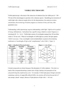
Transcription of Expt 2 Calcium by visible spectroscopy
1 1 | P a g e CHEM 450 expt . 2: DETERMINATION of Calcium in URINE SAMPLES by visible (Vis) spectroscopy Advanced Preparation: To be completed before coming to the lab. Show calculations to your instructor before preparing any of the solutions below. 100 mL of M Ca2+ from 400 ppm Ca2+ stock solution 200 mL of a x 10-3 M aqueous solution of Arsenazo III (FW g/mole) 1 L of M Imidazole buffer from the hydrochloride salt, adjusted to pH with either M HCl or M NaOH Background Soluble Calcium (as Ca2+ ions) in aqueous samples is usually determined by EDTA titration using Eriochrome Black T as indicator. In very dilute solutions, titration becomes impractical to use due to low levels of Calcium ions which make the color change at the equivalence point difficult to see. Calcium is usually measured to screen for or monitor diseases of the bone or Calcium regulation disorders (that is, diseases of the parathyroid gland or kidneys).
2 Urinary Calcium levels aid the clinician in understanding how the kidneys handle Calcium in certain disease states involving Calcium balance including diseases of the parathyroid gland. Urinary Calcium levels are also essential in the medical evaluation of kidney stones. ( ) Urine Calcium is usually measured in a sample taken from all the urine produced in a 24-hour period. Normal results may vary from lab to lab. Test results are affected by the amount of Calcium in the diet. Therefore, your health professional will consider the amount of Calcium in your diet when interpreting your urine Calcium levels. ( ) Calcium in urine Low amount of Calcium in diet: Less than 150 milligrams (mg)/24-hour sample Average amount of Calcium in diet: 100 250 mg/24-hour sample High amount of Calcium in diet: 250 300 mg/24-hour sample ultraviolet / visible (UV/vis) absorption spectroscopy is a technique that can be used to determine low levels of Calcium .
3 Because aqueous solutions of Calcium salts are colorless and therefore do not absorb in the near UV and visible region, an organic complexing agent will be used in this determination. Arsenazo III (1,8-dihydroxynaphthalene-3,6-disulphoni c acid-2,7-bis[(azo-2)phenylarsonic acid]) forms highly colored 1:1 complexes with Calcium and other metallic ions, allowing Calcium levels to be determined using UV/vis spectroscopy . Calcium -arsenazo complex is blue or purple in color, depending on pH, while uncomplexed arsenazo is wine-red. This complexation process is also utilized in the determination of Calcium ions in serum and urine. Calcium measurement in biological samples is important in the treatment of a variety of bone diseases and chronic renal disease. 2 | P a g e Ca2+ + Arsenazo III Ca2+ - Arsenazo III complex Colorless in water Wine-red Blue Because the complexation between Ca2+ ions and arsenazo is pH dependent, the solutions and samples will be buffered at pH Chemicals Used x 10-3 M Arsenazo III, disodium salt x 10-3 M Ca2+ from 400.
4 Ppm Ca2+ stock pH Imidazole buffer (see advanced prep) One 24-hr urine sample Procedure A. Preparation of Arsenazo and Calcium Chloride Solutions 1. (a) Using the FW of Arsenazo III ( g/mole for the disodium salt) calculate the amount in grams needed to prepare mL of a x 10-3 M aqueous Arsenazo III solution. Enter your calculation in your lab notebook. You do not have to weigh this. Instead, proceed to step 1(b). (b) Calculate the volume of 400. ppm Ca2+ stock solution needed to prepare mL of x 10-3 M Ca2+. Calculate the equivalent ppm concentration of this new dilution. Record both concentrations in your notebook. 2. For each group: Prepare mL of the x 10-3 M Ca2+ solution following your calculation above. The Arsenazo solution is already prepared based on the amount calculated above and is ready for use. B. Preparation of M pH Imidazole Buffer 1. Calibrate a pH meter using pH and buffers.
5 2. Calculate the amount of imidazole hydrochloride salt needed to prepare mL of M solution. Weigh the calculated amount on a weigh boat and transfer quantitatively (ask your instructor if you don t know what this means) into a 200-mL beaker. Dissolve in ~80mL of water. Check the pH of your buffer. Is it below or above the desired pH? 3. With constant stirring, add either M NaOH or M HCl dropwise until pH is reached. Transfer into a 100-mL volumetric flask. Dilute to the mark with DI water. Cap and shake well. 3 | P a g e C. Preparation of Calcium -Arsenazo III Standard Solutions and Unknown. 1. Obtain eight 25-mL volumetric flasks and label according to Table Flask ID. 2. Obtain one unknown urine sample from the hood for Calcium determination. Write down your unknown number. 3. Quantitatively transfer mL of your unknown into a mL volumetric flask. Dilute to the mark with deionized water.
6 (Save the remaining unknown sample for a future lab) 4. Label a 25-mL volumetric flask Patient (Unknown #) and another 25-mL volumetric flask 25-mL volumetric flask Patient (Unknown #) Duplicate . 5. Transfer mL unknown solution from step C3 into each of the labeled volumetric flasks using a calibrated micropipet. Do not dilute to the mark yet. Proceed to the next step. 6. Following Table prepare a solvent blank solution, four dilutions of the Ca-Arsenazo standard solution from part A buffered to pH (called working standards), a solution of Arsenazo III, and your unknown urine samples by combining the specified reagents into a volumetric flask. 7. Dilute each flask to the mark with deionized (DI) water. Cap and shake well. Allow at least 15 minutes for the complexation to take place before taking any absorbance measurements. 8. Using both the ppm and moles/L concentrations of your stock Ca-Arsenazo standard solution from part A, calculate the adjusted concentration of Ca2+ in the Ca-Arsenazo working standards following dilution with DI water to mL.
7 Enter these concentrations in the last two columns of Table [Hint: In all dilutions Ca2+ is the limiting reagent and Arsenazo III forms a 1:1 complex with Ca2+]. Table Amounts of reagents needed to prepare Ca2+- arsenazo III standard solutions. Flask ID Volume of x 10-3 M Arsenazo III (mL) Volume of x 10-3 M Ca2+ (mL) Volume of buffer (mL) Total Volume (mL) Concentration of Ca2+ in the working standard soln. (mol/L) Concentration of Ca2+ in the working standard soln. (ppm or mg/L) Blank 0 0 0 Arsenazo III 0 0 Std 1 Std 2 Std 3 Std 4 Patient __ 0 Patient __ duplicate 0 4 | P a g e D. Spectroscopic Determination of the Amount of Ca2+ in the Unknown Follow the operating instructions for the double-beam UV/Vis spectrophotometer. 1. Obtain the absorption spectrum of the blank solution. 2. Using the visible range of 450 to 750 nm wavelength obtain the absorption spectrum of your Arsenazo solution.
8 Compare this spectrum with that of the Ca-Arsenazo complex by overlapping the spectrum of Std 3 with that of Arsenazo. Print and label each spectrum accordingly. Question: From the result of the overlapped spectra, what wavelength should be used to quantify Ca2+ in the Ca-Arsenazo III complex and the unknowns? Why? 3. Depending on the display capability of the spectrophotometer, you may have to clear the absorption spectra obtained above before proceeding with the next steps. 4. Following the instructions on "Quantification" (also Quantitation) and using the wavelength of maximum absorption, max , generate a Calibration Curve by acquiring the spectra of the four working standards using an expanded wavelength range of 600 to 700 nm and plotting the resulting Absorbance vs. Concentration data. Select the option that displays the line equation and correlation coefficient. 5. Measure the absorbance of your unknown solution(s) and, using your calibration curve, calculate the concentration of Ca2+ in your unknown.
9 Remember to use the dilution factor (all your dilutions multiplied together) when calculating the concentration of Calcium ion in your original, undiluted unknown solution(s). 6. Print your data for inclusion in your laboratory report. Treatment and Reporting of Data 1. Calculate the mean ppm Ca2+, standard deviation, and % relative standard deviation (RSD) of duplicate measurements for your unknown. Report these in your RESULTS with the unknown number. 2. Is the [Ca2+] in your unknown normal or high? Discuss in enough details the implications of abnormal (both high and low) levels of Calcium in urine. Cite your references. Reference "Photometric Determination of Micro Amounts of Calcium with Arsenazo III." Anal. Chim. Acta, 53 (1971) 194-198.