Transcription of Doppler ultrasonography principles and methods of ...
1 F F F F Ferererererrrrrreireireireireira,a,a,a,a , F F F F Ign cio & C M Ign cio & C M Ign cio & C M Ign cio & C M Ign cio & C 2011. 2011. 2011. 2011. 2011. Doppler ultrasonography principles and methods of evaluation of thereproductive tract in mares. Acta Scientiae Veterinariae. 39(Supl 1): s105 - Scientiae Veterinariae, 2011. 39(Supl 1): s105 - Scientiae Veterinariae, 2011. 39(Supl 1): s105 - Scientiae Veterinariae, 2011. 39(Supl 1): s105 - Scientiae Veterinariae, 2011. 39(Supl 1): s105 - Scientiae Veterinariae, 2011. 39(Supl 1): s105 - 1679-9216 (Online)1 Programa de P s-gradua o em Medicina Veterin ria, FMVZ, Departamento de Reprodu o Animal e Radiologia Veterin ria, UniversidadeEstadual Paulista J lio de Mesquita Filho, Botucatu, SP, Brasil. 2 Faculdade de Medicina Veterin ria e Zootecnia, Universidade Estadual PaulistaJ lio de Mesquita Filho, Departamento de Reprodu o Animal e Radiologia Veterin ria, Botucatu, SP, Brasil.
2 CORRESPOND NCIA: Departamento de Reprodu o Animal e Radiologia Veterin ria, Universidade Estadual Paulista J lio deMesquita Filho. CEP18608-210 Botucatu, SP, ultrasonography principles and methods of evaluation of theDoppler ultrasonography principles and methods of evaluation of theDoppler ultrasonography principles and methods of evaluation of theDoppler ultrasonography principles and methods of evaluation of theDoppler ultrasonography principles and methods of evaluation of thereproductive tract in maresreproductive tract in maresreproductive tract in maresreproductive tract in maresreproductive tract in maresJair Camargo FerreiraJair Camargo FerreiraJair Camargo FerreiraJair Camargo FerreiraJair Camargo Ferreira11111, Fernanda Saules Ign cio, Fernanda Saules Ign cio, Fernanda Saules Ign cio, Fernanda Saules Ign cio, Fernanda Saules Ign cio22222 & Cezinande de Meira & Cezinande de Meira & Cezinande de Meira & Cezinande de Meira & Cezinande de Meira22222 ABSTRACTB ackground: Doppler ultrasonography is a non-invasive real time pulse-wave technique recently used for the transrectalstudy of the reproductive system hemodynamics in large animals.
3 This technic is based in the Doppler Effect Principle thatproposes the change in frequency of a wave for an observer (red blood cells) moving relative to the source of the respectivewave (ultrasonic transducer). This method had showed to be effective and useful for the evaluation of the in vivo equinereproductive tract increasing the diagnostic, monitoring, and predictive capabilities of theriogenology in mares. However, anaccurate and truthful ultrasonic exam requires the previous knowledge of the Doppler ultrasonography : In recent years, the capabilities of ultrasound flow imaging have increased enormously. The current Doppler ultrasoundmachines offer three methods of evaluation that may be used simultaneously (triplex mode). In B-mode ultrasound, a lineararray of transducers simultaneously scans a plane through the tissue that can be viewed as a two-dimensional gray-scale imageon screen. This mode is primarily used to identify anatomically a structure for its posterior evaluation using colored ultrasoundmodes (Color or Spectral modes).
4 Colored ultrasound images of flow, whether Color or Spectral modes, are essentiallyobtained from measurements of moving red cells. In Color mode, velocity information is presented as a color coded overlay ontop of a B-mode image, while Pulsed Wave Doppler provides a measure of the changing velocity throughout the cardiac cycleand the distribution of velocities in the sample volume represented by a spectral graphic. Color images conception variesaccording to the Doppler Frequency that is the difference between the frequency of received echoes by moving blood red cellsand wave frequency transmitted by the transducer. To produce an adequate spectral graphic it is important determine theposition and size of the simple gate. Furthermore, blood flow velocity measurement is influence by the intersection anglebetween ultrasonic pulses and the direction of moving blood-red cells ( Doppler angle).
5 Objectively colored ultrasound exammay be done on large arteries of the reproductive tract, as uterine and ovary arteries, or directly on the target tissue (follicle, forexample). Mesovarium and mesometrium attachment arteries also can be used for spectral evaluation of the equine reproductivesystem. Subjectively analysis of the ovarian and uterine vascular perfusion must be done directly on the corpus luteum,follicular wall and uterus (endometrium and myometrium associated), respectively. Power- flow imaging has greater sensitivityto weak blood flow and independent of the Doppler angle, improving the evaluation of vessels with small diameters and slowblood : Doppler ultrasonography principles , methods of evaluation and reproductive system anatomy have been knowledge is essential for the competent equipment acquisition and precise collection and analysis of colored ultrasoundimages. Otherwise, the reporting of inconsistent and not reproducible findings may result in the discredit of Doppler technologyahead of the scientific veterinary : Doppler ultrasound imaging , hemodynamics, blood-red cells, spectrum, color-flow, F F F F Ferererererrrrrreireireireireira,a,a,a,a , F F F F Ign cio & C M Ign cio & C M Ign cio & C M Ign cio & C M Ign cio & C 2011.
6 2011. 2011. 2011. 2011. Doppler ultrasonography principles and methods of evaluation of thereproductive tract in mares. Acta Scientiae Veterinariae. 39(Supl 1): s105 - INTRODUCTIONII. COLORED ULTRASONIC Spectral Color-flow modeIII. ARTERIAL SYSTEM OF THE REPRODUCTIVETRACTIV. CONCLUSIONI. INTRODUCTIONIn large animals, Doppler ultrasonography isa non-invasive real time pulse-wave technique recen-tly used for the transrectal study of the reproductivesystem hemodynamics. The introduction of thistechnology in current researches has allowedreevaluating previously conceptions considereddefinitive regarding the physiology of method had showed to be effective and usefulfor the evaluation of the in vivo equine reproductivetract increasing the diagnostic, monitoring, andpredictive capabilities of theriogenology in mares [4].
7 Color- Doppler ultrasonic imaging is based inthe Doppler effect principle that proposes the changein frequency of a wave for an observer movingrelative to the source of the respective wave (Figure1). The frequency is constant when the wave sourceand observer are stationary. However, if there is amoving toward or away from each other, the returningechoes frequency increase and decrease, respectively[7]. At the moment, this principle is used for differentapplications as temperature, velocity and vibrationmeasurements. In Doppler sonography, the wavesource and stationary object are, respectively, the red-blood cells and the an ultrasonic Doppler exam is possibleto perform a qualitatively and/or quantitativelyevaluation of specific vessels and tissues. Thedirection and velocity of red-blood cells can berepresented by different types and intensities of colorspixels or as a velocity spectral graphic.
8 However, toselect the most appropriated mode of exam first it isnecessary to have a complete knowledge of theDoppler ultrasonography principles and methods COLORED ULTRASONIC IMAGINGT here are two specifically approaches forcolored ultrasonic imaging evaluation: Color-flow andSpectral modes (Figure 2). Color-flow mode usesColor- Doppler signals superimposes on a B-modeFigure 1. Collection of images representing the Doppler Effect. Wave frequency emitted by a staticobject (Vsource = 0) relative to the observer is constant, concentric and equally spaced. When the pulseis emitted during relative motion between source and observer, the wave fronts are no longer frequency of echoes increases and decreases, respectively, during approach and away movements(Vsource < Vsound). When the velocity of the object is similar to the sound speed (Vsource =Vsound), the crests of waves emitted overlap forming a single ridge that reaches the observersimultaneously with the source.
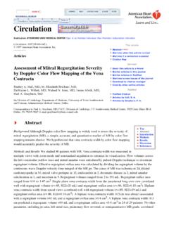
9 Emitted wave reaches the observer after passing the sound source atsupersonic speeds (Vsource > Vsom). Font: Personal 2. Ultrasonic images collection of female reproductive tract in B, Power, Color and Spectral modes (A, B, C and D, respectively). (A andB) Cross section image, using B-mode and Power-flow Doppler , of a non-pregnant uterine horn in diestrus. (C) Color Doppler evaluation ofa pre-ovulatory follicle. (B and C) Presence of colored pixels within uterine tissue (endometrium and myometrium) and follicularwall indicate the red blood cells displacement and have been used to estimate the tissue vascularity. (D) Spectral graphconsisting of maximum speeds values of blood flow in mesometrial artery. Source: Personal F F F F Ferererererrrrrreireireireireira,a,a,a,a , F F F F Ign cio & C M Ign cio & C M Ign cio & C M Ign cio & C M Ign cio & C 2011. 2011. 2011. 2011. 2011. Doppler ultrasonography principles and methods of evaluation of thereproductive tract in mares.
10 Acta Scientiae Veterinariae. 39(Supl 1): s105 - of a structure to estimate its vascularity, whilepulsed- Doppler spectral analysis of blood velocitiesof a specific artery is done on Spectral mode. Bothmethods are based on Doppler -shift frequencies, alsocalled Doppler frequency [7]. Doppler frequency is the difference betweenthe frequency of received echoes and frequencytransmitted by the transducer (Figure 3). Thetransducer transmitted frequency is constant whilethe frequency of the returning echoes varies accordingto the Doppler Effect principle. Therefore, when thewave source (blood-red cells) is stationary or movingparallel to the transducer there is no differencebetween transmitted and returning frequencies andcolored Doppler signals are not detected. If the bloodflow moves toward to the transducer, the returningfrequency is greater than the transmitted frequencyresulting in a positive Doppler frequency.